

Midline suboccipital approach to a vertebral artery-posterior inferior cerebellar artery aneurysm from the rostral end of the patient using ORBEYE
- PMID: 35399900
- PMCID: PMC8986755
- DOI: 10.25259/SNI_1272_2021
Midline suboccipital approach to a vertebral artery-posterior inferior cerebellar artery aneurysm from the rostral end of the patient using ORBEYE
Abstract
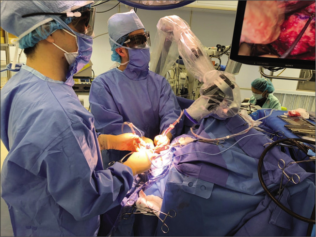
Background: The midline suboccipital approach with the patient in the prone position is safe and effective for clipping vertebral artery-posterior inferior cerebellar artery (VA-PICA) aneurysms. Using a conventional surgical microscope from the rostral end of the patient for this approach without an extreme head-down position requires the surgeon to overhang the visual axis of the microscope and perform surgical manipulations in an uncomfortable posture. We report performing the midline suboccipital approach from the rostral end with slight head-down position using ORBEYE, a new high-definition (4K) three-dimensional exoscope.
Case description: A 65-year-old woman was admitted for clipping of a right unruptured VA-PICA aneurysm (maximum diameter, 5mm) located medially and ventral to the hypoglossal canal. After induction of general anesthesia, the patient was placed in the prone position with the head titled slightly downward. A midline suboccipital approach was performed from the rostral end of the patient using ORBEYE. Clipping was safely accomplished in a comfortable posture. No operative complications occurred. Postoperative computed tomography angiography showed complete aneurysmal obstruction.
Conclusion: Exoscopic surgery using ORBEYE is feasible for a midline suboccipital approach to VA-PICA aneurysms from the rostral end of the patient with the patient in the prone with slight head-down position.
Keywords: Clipping; Exoscope; Midline suboccipital approach; ORBEYE; Posterior inferior cerebellar artery aneurysm.
Copyright: © 2022 Surgical Neurology International.
Conflict of interest statement
There are no conflicts of interest.
Figures


Similar articles
-
[Contralateral suboccipital approach for clipping of an unruptured vertebral artery-posterior inferior cerebellar artery aneurysm].No Shinkei Geka. 2014 Jan;42(1):35-40. No Shinkei Geka. 2014. PMID: 24388938 Review. Japanese.
-
[Surgery of vertebral artery aneurysm at the origin of posterior inferior cerebellar artery--with special reference to operative technics in cases with difficulty in direct operation].No Shinkei Geka. 1984 Jul;12(8):933-41. No Shinkei Geka. 1984. PMID: 6483100 Japanese.
-
Aneurysms of the posterior inferior cerebellar artery-vertebral artery complex: variations on a theme.Neurosurgery. 1990 Jul;27(1):12-20; discussion 20-1. doi: 10.1097/00006123-199007000-00002. Neurosurgery. 1990. PMID: 2377268
-
Utility of a novel exoscope, ORBEYE, in gravity-assisted brain retraction surgery for midline lesions of the brain.Surg Neurol Int. 2021 Jul 6;12:339. doi: 10.25259/SNI_320_2021. eCollection 2021. Surg Neurol Int. 2021. PMID: 34345480 Free PMC article.
-
[Multiple aneurysms of the distal posterior inferior cerebellar artery with recurrent hemorrhage undetectable on preoperative neuroradiological findings: case report].No Shinkei Geka. 2004 Nov;32(11):1157-64. No Shinkei Geka. 2004. PMID: 15570881 Review. Japanese.
Cited by
-
The Usefulness of Surgical Titanium Microclips for Mucosal Repair in the Frontal Sinus Using ORBEYE: A Technical Note.Neurol Med Chir (Tokyo). 2024 Mar 15;64(3):131-135. doi: 10.2176/jns-nmc.2023-0175. Epub 2024 Jan 31. Neurol Med Chir (Tokyo). 2024. PMID: 38296551 Free PMC article.
-
Microsurgical Resection of Meningiomas Using a 4K Three-Dimensional Exoscope: A Descriptive Observational Study.Cureus. 2024 Dec 2;16(12):e74950. doi: 10.7759/cureus.74950. eCollection 2024 Dec. Cureus. 2024. PMID: 39749091 Free PMC article.
-
High-definition two-dimension video telescope operating monitor-assisted brain and spinal surgery in pediatrics: is it an acceptable substitute for microscopic surgery?Childs Nerv Syst. 2022 Nov;38(11):2171-2177. doi: 10.1007/s00381-022-05636-y. Epub 2022 Aug 9. Childs Nerv Syst. 2022. PMID: 35943568
References
-
- Al-Khayat H, Al-Khayat H, Beshay J, Manner D, White J. Vertebral artery-posteroinferior cerebellar artery aneurysms: Clinical and lower cranial nerve outcomes in 52 patients. Neurosurgery. 2005;56:2–10. discussion 11. - PubMed
-
- Chalouhi N, Jabbour P, Starke RM, Tjoumakaris SI, Gonzalez LF, Witte S, et al. Endovascular treatment of proximal and distal posterior inferior cerebellar artery aneurysms. J Neurosurg. 2013;118:991–9. - PubMed
-
- Forget TR, Jr, Benitez R, Veznedaroglu E, Sharan A, Mitchell W, Silva M, et al. A review of size and location of ruptured intracranial aneurysms. Neurosurgery. 2001;49:1322–5. discussion 1325-6. - PubMed
Publication types
LinkOut - more resources
Full Text Sources