

Visual Counting and Automated Image-analytic Assessment of Ki-67 and their Prognostic Value in Synovial Sarcoma
- PMID: 35400010
- PMCID: PMC8962852
- DOI: 10.21873/cdp.10070
Visual Counting and Automated Image-analytic Assessment of Ki-67 and their Prognostic Value in Synovial Sarcoma
Abstract
Background: Ki-67 is a widely used proliferation marker reflecting prognosis in various tumors. However, visual assessment and scoring of Ki-67 suffers from marked inter-observer and intra-observer variability. We aimed to assess the concordance of manual counting and automated image-analytic scoring methods for Ki-67 in synovial sarcoma.
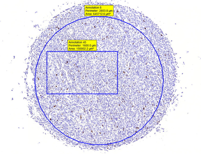
Patients and methods: Tissue microarrays from 34 patients with synovial sarcoma were immunostained for Ki-67 and scored both visually and with 3DHistech QuantCenter.
Results: The automated assessment of Ki-67 expression was in good agreement with the visually counted Ki-67 (r Pearson =0.96, p<0.001). In a Cox regression model automated [hazard ratio (HR)=1.047, p=0.024], but not visual (HR=1.063, p=0.053) assessment method associated high Ki-67 scores with worse overall survival.
Conclusion: The automated Ki-67 assessment method appears to be comparable to the visual method in synovial sarcoma and had a significant association to overall survival.
Keywords: Ki-67; Ki-67 antigen; Sarcoma; automated; pattern recognition; survival analysis; synovial; visual counting method.
Copyright 2022, International Institute of Anticancer Research.
Conflict of interest statement
The Authors declare that there are no conflicts of interest regarding the publication of this paper.
Figures


References
-
- Ladanyi M, Antonescu CR, Leung DH, Woodruff JM, Kawai A, Healey JH, Brennan MF, Bridge JA, Neff JR, Barr FG, Goldsmith JD, Brooks JS, Goldblum JR, Ali SZ, Shipley J, Cooper CS, Fisher C, Skytting B, Larsson O. Impact of SYT-SSX fusion type on the clinical behavior of synovial sarcoma: a multi-institutional retrospective study of 243 patients. Cancer Res. 2002;62(1):135–140. - PubMed
-
- Gerdes J, Lemke H, Baisch H, Wacker HH, Schwab U, Stein H. Cell cycle analysis of a cell proliferation-associated human nuclear antigen defined by the monoclonal antibody Ki-67. J Immunol. 1984;133(4):1710–1715. - PubMed
-
- Ogino J, Asanuma H, Hatanaka Y, Matsuno Y, Gotoda H, Muraoka S, Tsuji T, Fukazawa Y, Yamashiro K, Kondo N, Iwaki H, Miyokawa N, Hasegawa T. Validity and reproducibility of Ki-67 assessment in gastrointestinal stromal tumors and leiomyosarcomas. Pathol Int. 2013;63(2):102–107. doi: 10.1111/pin.12038. - DOI - PubMed
LinkOut - more resources
Full Text Sources