Effects of Prestretch on Neonatal Peripheral Nerve: An In Vitro Study
- PMID: 35400085
- PMCID: PMC8993512
- DOI: 10.1055/s-0042-1743132
Effects of Prestretch on Neonatal Peripheral Nerve: An In Vitro Study
Abstract
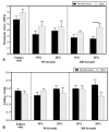
Background Characterizing the biomechanical failure responses of neonatal peripheral nerves is critical in understanding stretch-related peripheral nerve injury mechanisms in neonates. Objective This in vitro study investigated the effects of prestretch magnitude and duration on the biomechanical failure behavior of neonatal piglet brachial plexus (BP) and tibial nerves. Methods BP and tibial nerves from 32 neonatal piglets were harvested and prestretched to 0, 10, or 20% strain for 90 or 300 seconds. These prestretched samples were then subjected to tensile loading until failure. Failure stress and strain were calculated from the obtained load-displacement data. Results Prestretch magnitude significantly affected failure stress but not the failure strain. BP nerves prestretched to 10 or 20% strain, exhibiting significantly lower failure stress than those prestretched to 0% strain for both prestretch durations (90 and 300 seconds). Likewise, tibial nerves prestretched to 10 or 20% strain for 300 seconds, exhibiting significantly lower failure stress than the 0% prestretch group. An effect of prestretch duration on failure stress was also observed in the BP nerves when subjected to 20% prestretch strain such that the failure stress was significantly lower for 300 seconds group than 90 seconds group. No significant differences in the failure strains were observed. When comparing BP and tibial nerve failure responses, significantly higher failure stress was reported in tibial nerve prestretched to 20% strain for 300 seconds than BP nerve. Conclusion These data suggest that neonatal peripheral nerves exhibit lower injury thresholds with increasing prestretch magnitude and duration while exhibiting regional differences.
Keywords: brachial plexus; injury; neonates; prestretch; stretch; tibial nerve.
The Author(s). This is an open access article published by Thieme under the terms of the Creative Commons Attribution License, permitting unrestricted use, distribution, and reproduction so long as the original work is properly cited. ( https://creativecommons.org/licenses/by/4.0/ ).
Conflict of interest statement
Conflict of Interest None declared.
Figures


References
-
- Rydevik B L, Kwan M K, Myers R R. An in vitro mechanical and histological study of acute stretching on rabbit tibial nerve. J Orthop Res. 1990;8(05):694–701. - PubMed
-
- Kwan M K, Wall E J, Weiss J A, Rydevik B L, Garfin S R, Woo S-Y. Analysis of rabbit peripheral nerve: in situ stresses and strains. Biomech Symp ASME/AMD. 1989;98:109–112.
-
- Li J, Shi R. Stretch-induced nerve conduction deficits in guinea pig ex vivo nerve. J Biomech. 2007;40(03):569–578. - PubMed
-
- Bain A C, Raghupathi R, Meaney D F. Dynamic stretch correlates to both morphological abnormalities and electrophysiological impairment in a model of traumatic axonal injury. J Neurotrauma. 2001;18(05):499–511. - PubMed
-
- Sunderland S. The anatomy and physiology of nerve injury. Muscle Nerve. 1990;13(09):771–784. - PubMed
LinkOut - more resources
Full Text Sources

