. 2022 Mar 24:16:816174.
doi: 10.3389/fnins.2022.816174. eCollection 2022.

Mitigating Effect of Estrogen in Alzheimer's Disease-Mimicking Cerebral Organoid

Affiliations

Mitigating Effect of Estrogen in Alzheimer's Disease-Mimicking Cerebral Organoid

Jennifer Yejean Kim et al. Front Neurosci. .

Abstract

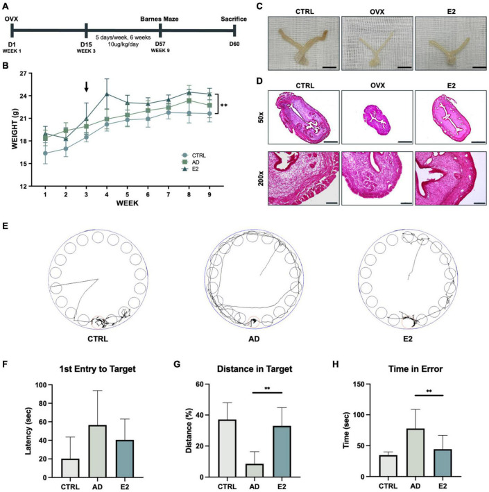
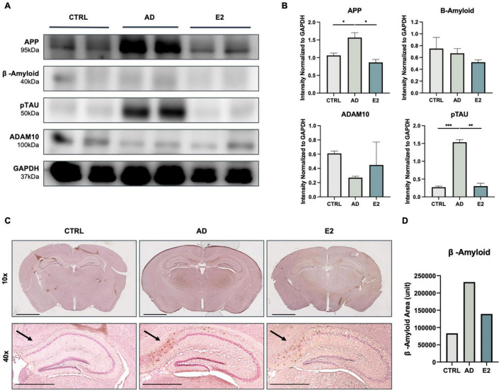
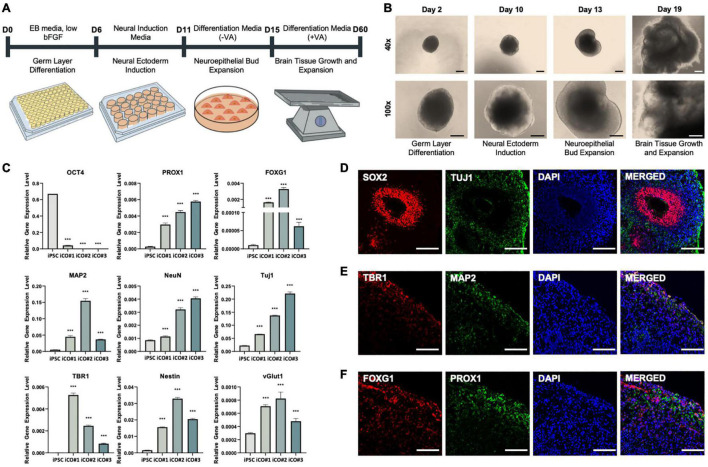
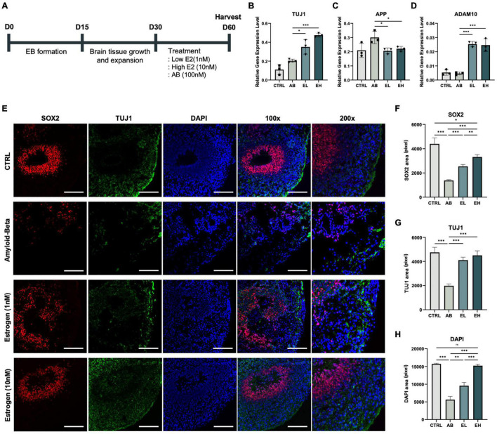
Alzheimer's disease (AD) is the most common condition in patients with dementia and affects a large population worldwide. The incidence of AD is expected to increase in future owing to the rapid expansion of the aged population globally. Researchers have shown that women are twice more likely to be affected by AD than men. This phenomenon has been attributed to the postmenopausal state, during which the level of estrogen declines significantly. Estrogen is known to alleviate neurotoxicity in the brain and protect neurons. While the effects of estrogen have been investigated in AD models, to our knowledge, they have not been investigated in a stem cell-based three-dimensional in vitro system. Here, we designed a new model for AD using induced pluripotent stem cells (iPSCs) in a three-dimensional, in vitro culture system. We used 5xFAD mice to confirm the potential of estrogen in alleviating the effects of AD pathogenesis. Next, we confirmed a similar trend in an AD model developed using iPSC-derived cerebral organoids, in which the key characteristics of AD were recapitulated. The findings emphasized the potential of estrogen as a treatment agent for AD and also showed the suitability of AD-recapitulating cerebral organoids as a reliable platform for disease modeling and drug screening.

Keywords: Alzheimer’s disease; amyloid-beta; cerebral organoid; estrogen; induced pluripotent stem cells.


Conflict of interest statement

JK and YN are employed by YiPSCELL, Inc. JHJ is founder of YiPSCELL, Inc. The remaining authors declare that the research was conducted in the absence of any commercial or financial relationships that could be construed as a potential conflict of interest.

Figures

FIGURE 1
FIGURE 1
Estrogen treatment of an Alzheimer’s disease model developed using ovariectomized mice improves behavioral performance. (A) Schematic representation of the animal experiment, involving the induction of the postmenopausal state in the Alzheimer’s disease model of mice and treatment with estrogen. (B) Growth curves of control mice and ovariectomized (OVX) mice treated with estrogen. Arrows indicate the time of OVX operation and the starting point of estrogen injection. (C) Uteri of control (C57BL/6), OVX, and OVX + E2 mice. Scale bar, 1 cm. (D) Hematoxylin and eosin staining of mice uterine tissues. Scale bar, 500 μm for 50 × ; 100 μm for 200 ×. (E) Monitoring of mice from each group for 3 min (180 s). (F) Graph showing the average latency of first entry to target in seconds. (G) Graph showing the average distance traveled in the target relatively. (H) Graph showing the average time spent by the mice outside the target zone in seconds. t-tests were used for data analysis (**P < 0.01). Values are presented as mean ± standard error of the mean.
FIGURE 2
FIGURE 2
Histological analyses of brain tissues from control, 5xFAD, and ovariectomized 5xFAD model mice treated with estrogen. (A) Western blotting for the measurement of amyloid precursor protein (APP), amyloid-beta (Aβ) protein, phosphorylated tau (TAU) protein, and ADAM10 protein levels in each group. (B) Quantification in western blotting assay with protein levels normalized to that of GAPDH. (C) Immunohistochemical assay showed Aβ plaque accumulation in mouse brains, with significant clustering in the cortical and hippocampal regions. Scale bar, 2000 μm for 10x, 1000 μm for 40x. (D) Quantification of Aβ-positive areas, measured using ImageJ. t-tests were used for data analysis (*P < 0.05, **P < 0.01, ***P < 0.001).
FIGURE 3
FIGURE 3
Successful differentiation of cerebral organoids from induced pluripotent stem cells obtained from healthy patient. (A) Differentiation of cerebral organoids derived from induced pluripotent stem cells. (B) Images of cerebral organoids in each step of the experiment. (C) Gene expression normalized to that of GAPDH: pluripotency marker OCT4, hippocampal marker PROX1, forebrain marker FOXG1, neuronal markers MAP2, NeuN, and TUJ1, deep-layer neuron marker TBR1, neural progenitor cell marker Nestin, and glutamate transporter marker vGLUT1. Immunofluorescence assay showing the co-localization of (D) early-stage neuronal marker SOX2 and neuronal cell-body marker TUJ1, (E) deep-layer neuron marker TBR1 and neuronal dendrite marker MAP2, and (F) forebrain marker FOXG1 and hippocampal marker PROX1, in the cerebral organoids. t-tests were performed for data analysis (***P < 0.001). Values are presented as mean ± standard error of mean. Scale bars, 200 μm.
FIGURE 4
FIGURE 4
Estrogen treatment of cerebral organoids co-stimulated with amyloid-beta (Aβ) enhances the expression of neuronal markers. (A) Schematic representation of the cerebral organoid generation process and treatment with Aβ peptide and estrogen. Gene expression normalized to that of GAPDH: (B) neuronal markers TUJ1, (C) amyloid precursor protein marker APP, and (D) alpha-secretase marker ADAM10. (E) Immunofluorescence assay showing the co-localization of early-stage neuronal marker SOX2 and neuronal markers TUJ1 in samples obtained from experimental and control mice. (F-H) Areas that stained positive for DAPI, SOX2, and TUJ1 were measured using ImageJ. CTRL, control; Aβ, cerebral organoids treated only with 100 nM Aβ; EL, cerebral organoids treated with 100 nM Aβ and 1 nM estrogen; EH, cerebral organoids treated with 100 nM Aβ and 10 nM estrogen. t-tests were performed for data analysis (*P < 0.05, **P < 0.01, ***P < 0.001). Values are presented as mean ± standard error of mean. Scale bars, 200 μm.

Similar articles

Cited by

References

    1. Allinson T. M., Parkin E. T., Turner A. J., Hooper N. M. (2003). ADAMs family members as amyloid precursor protein α−secretases. J. Neurosci. Res. 74 342–352. 10.1002/jnr.10737 - DOI - PubMed
    1. Alzamora R., Brown L. R., Harvey B. J. (2007). Direct binding and activation of protein kinase C isoforms by aldosterone and 17β-estradiol. Mole. Endocrinol. 21 2637–2650. - PubMed
    1. Amtul Z., Wang L., Westaway D., Rozmahel R. F. (2010). Neuroprotective mechanism conferred by 17beta-estradiol on the biochemical basis of Alzheimer’s disease. Neuroscience 169 781–786. 10.1016/j.neuroscience.2010.05.031 - DOI - PubMed
    1. Anastasio T. J. (2013). Exploring the contribution of estrogen to amyloid-Beta regulation: a novel multifactorial computational modeling approach. Front. Pharmacol. 4:16. 10.3389/fphar.2013.00016 - DOI - PMC - PubMed
    1. Andorfer C., Kress Y., Espinoza M., de Silva R., Tucker K. L., Barde Y. A., et al. (2003). Hyperphosphorylation and aggregation of tau in mice expressing normal human tau isoforms. J. Neurochem. 86 582–590. 10.1046/j.1471-4159.2003.01879.x - DOI - PubMed

LinkOut - more resources