

Expression of SH3 and Multiple Ankyrin Repeat Domains Protein 3 in Mouse Retina
- PMID: 35401120
- PMCID: PMC8990853
- DOI: 10.3389/fncel.2022.795668
Expression of SH3 and Multiple Ankyrin Repeat Domains Protein 3 in Mouse Retina
Abstract
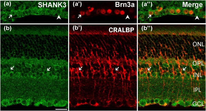
Synapse-associated gene mutations of SH3 and multiple ankyrin repeat domains protein 3 (SHANK3) may lead to autism spectrum disorder (ASD). In some ASD cases, patients may also have vision disorders. However, the effects of SHANK3 in the retina are barely mentioned in the literature. In this study, we used wild-type mice to systematically map the distribution of SHANK3 expression in entire mouse retinas. Using Western blot analysis and immunofluorescence double labeling, we identified a large number of prominent cells expressing high levels of SHANK3 in the inner retina, in particular, the ganglion cell layer (GCL) and inner nucleus layer. The inner plexiform layer and outer nucleus layer were also exhibited positive SHANK3 signals. In the inner layer, GABAergic amacrine cells (ACs) labeled by glutamate decarboxylase were colocalized with SHANK3-positive cells. Dopaminergic ACs (labeled by tyrosine hydroxylase) and cholinergic ACs (labeled by choline acetyltransferase) were also found to contain SHANK3-positive signals. Additionally, most GCs (labeled by Brn3a) were also found to be SHANK3 positive. In the outer retina, bipolar cells (labeled by homeobox protein ChX10) and horizontal cells (labeled by calbindin) were SHANK3 positive. In the outer nucleus layers, the somata of blue cones (labeled by S-opsin) were weekly co-labeled with SHANK3. The inner segments of blue cones and the outer segments of red/green cones (labeled by L/M-opsin) were partially colocalized with SHANK3 and the outer segments of rods (labeled by Rho4D2) were partially SHANK3 positive too. Moreover, SHANK3-positive labeling was also observed in Müller cells (labeled by cellular retinaldehyde-binding protein). These wide expression patterns indicate that SHANK3 may play an important role in the visual signaling pathway.
Keywords: ASD; SHANK3; double-labeled immunohistochemistry; excitatory synapse; retina.
Copyright © 2022 Xu, Wang, Tong, Li, Cheng, Zhang, Xu, Wang and Zhang.
Conflict of interest statement
The authors declare that the research was conducted in the absence of any commercial or financial relationships that could be construed as a potential conflict of interest.
Figures


Similar articles
-
Cellular localization of the FMRP in rat retina.Biosci Rep. 2020 Jun 26;40(6):BSR20200570. doi: 10.1042/BSR20200570. Biosci Rep. 2020. PMID: 32452512 Free PMC article.
-
Cellular localization of P2Y₆ receptor in rat retina.Neuroscience. 2012 Sep 18;220:62-9. doi: 10.1016/j.neuroscience.2012.06.032. Epub 2012 Jun 21. Neuroscience. 2012. PMID: 22728100
-
Gene expression and protein distribution of orexins and orexin receptors in rat retina.Neuroscience. 2011 Aug 25;189:146-55. doi: 10.1016/j.neuroscience.2011.04.011. Epub 2011 May 27. Neuroscience. 2011. PMID: 21621592
-
Localization of neuropeptide receptor NPY4R in rat retina.Neuropeptides. 2022 Jun;93:102246. doi: 10.1016/j.npep.2022.102246. Epub 2022 Apr 9. Neuropeptides. 2022. PMID: 35453028
-
Targeting Shank3 deficiency and paresthesia in autism spectrum disorder: A brief review.Front Mol Neurosci. 2023 Feb 9;16:1128974. doi: 10.3389/fnmol.2023.1128974. eCollection 2023. Front Mol Neurosci. 2023. PMID: 36846568 Free PMC article. Review.
Cited by
-
Elevated SH3 and Multiple Ankyrin Repeat Domains 2 Expression Correlates With Improved Glioma Prognosis.Int J Genomics. 2024 Oct 4;2024:6565925. doi: 10.1155/2024/6565925. eCollection 2024. Int J Genomics. 2024. PMID: 39397895 Free PMC article.
References
LinkOut - more resources
Full Text Sources
Molecular Biology Databases
Research Materials