. 2022 Mar 24:13:845018.
doi: 10.3389/fphar.2022.845018. eCollection 2022.

Three-Day Continuous Oxytocin Infusion Attenuates Thermal and Mechanical Nociception by Rescuing Neuronal Chloride Homeostasis via Upregulation KCC2 Expression and Function

Affiliations

Three-Day Continuous Oxytocin Infusion Attenuates Thermal and Mechanical Nociception by Rescuing Neuronal Chloride Homeostasis via Upregulation KCC2 Expression and Function

Xiyuan Ba et al. Front Pharmacol. .

Abstract

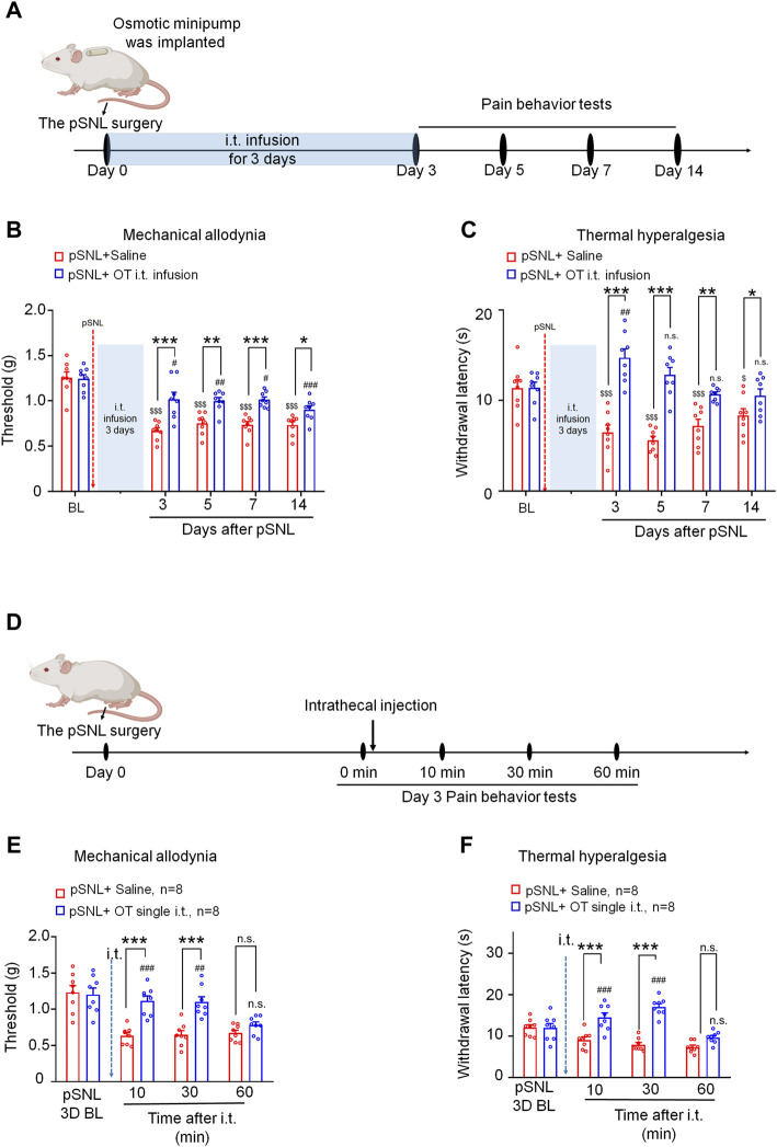
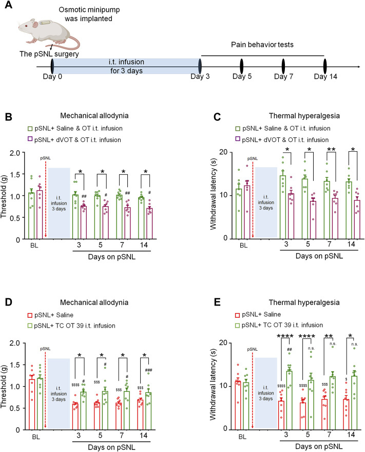
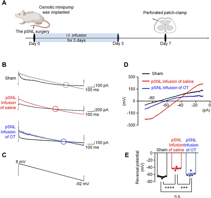
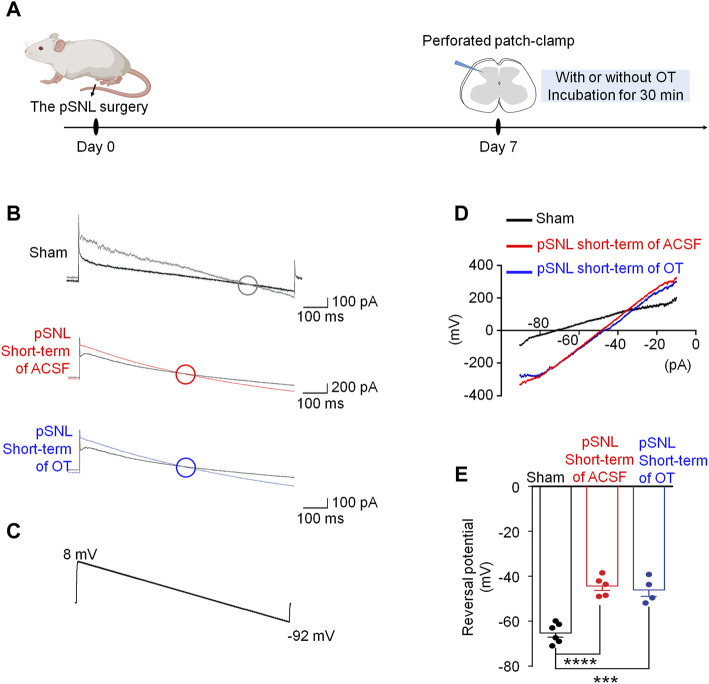
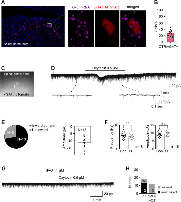
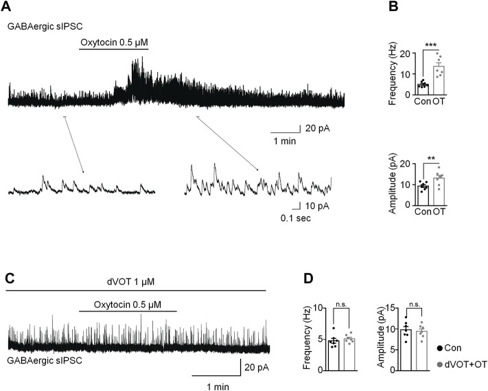
Oxytocin (OT) and its receptor are promising targets for the treatment and prevention of the neuropathic pain. In the present study, we compared the effects of a single and continuous intrathecal infusion of OT on nerve injury-induced neuropathic pain behaviours in mice and further explore the mechanisms underlying their analgesic properties. We found that three days of continuous intrathecal OT infusion alleviated subsequent pain behaviours for 14 days, whereas a single OT injection induced a transient analgesia for 30 min, suggesting that only continuous intrathecal OT attenuated the establishment and development of neuropathic pain behaviours. Supporting this behavioural finding, continuous intrathecal infusion, but not short-term incubation of OT, reversed the nerve injury-induced depolarizing shift in Cl- reversal potential via restoring the function and expression of spinal K+-Cl- cotransporter 2 (KCC2), which may be caused by OT-induced enhancement of GABA inhibitory transmission. This result suggests that only continuous use of OT may reverse the pathological changes caused by nerve injury, thereby mechanistically blocking the establishment and development of pain. These findings provide novel evidence relevant for advancing understanding of the effects of continuous OT administration on the pathophysiology of pain.

Keywords: K+-Cl-cotransporter 2; chloride homeostasis; continuous intrathecal drug delivery; neuropathic pain; oxytocin.


Conflict of interest statement

The authors declare that the research was conducted in the absence of any commercial or financial relationships that could be construed as a potential conflict of interest.

Figures

FIGURE 1
FIGURE 1
Three-day continuous intrathecal infusion, but not short-term application of OT, attenuated the establishment and development of nerve injury-induced nociceptive behaviours in pSNL mice. (A) A schematic of the experimental design. (B,C) Continuous intrathecal OT infusion (0.3 μg/100 μL) for 3 days before behavioural tests decreased pSNL-induced mechanical allodynia (A) and thermal hyperalgesia (B) for 14 days. (D) A schematic of the experimental design. (E,F) A single intrathecal OT injection (0.1 μg/10 μL) relieved pSNL-induced mechanical allodynia (E) and thermal hyperalgesia (F) in mice. Two-way repeated-measures ANOVA with group as the between-subjects factor and day/time as the within-subjects factor. Data are expressed as mean ± SEM. *p < 0.05, **p < 0.01, ***p < 0.001 OT vs. saline; $ p < 0.05, $$$ p < 0.001 vs. baseline; # p < 0.05, ## p < 0.01, #### p < 0.0001 vs. baseline.
FIGURE 2
FIGURE 2
The effects of 3-days OT infusion on nerve injury-induced nociceptive behaviours were mediated by OXTRs. (A) A schematic of the experimental design. (B,C) OT’s effect on mechanical allodynia (B) and thermal hyperalgesia (C) was completely blocked by its selective antagonist, dVOT (0.3 μg/100 μL). (D,E) Selective OT receptor agonists, TC OT (0.3 μg/100 μL, intrathecal infusion) showed similar effects on mechanical allodynia (D) and thermal hyperalgesia (E) in pSNL mice. Two-way repeated-measures ANOVA with group as the between-subjects factor. Data are expressed as mean ± SEM. *p < 0.05, **p < 0.01, ***p < 0.001 TC OT vs. saline; OT vs. dVOT and OT. $$$ p < 0.001, $$$$ p < 0.0001 vs. baseline; # p < 0.05, ## p < 0.01, ### p < 0.001 vs. baseline.
FIGURE 3
FIGURE 3
Three-day continuous intrathecal OT infusion renormalized EGABA in spinal dorsal horn. (A) The schematics of the electrophysiological recording. (B,C) As voltage ramps applied from +8 to −92 Mv (C), basal and GABA-evoked currents were recorded (B). (D,E) Representative (D) and statistical (E) reversal potential of EGABA recorded from slices of sham and pSNL mice treated with continuous OT or saline. One-way ANOVA followed by Bonferroni’s post hoc test. Data are expressed as mean ± SEM. ***p < 0.001 sham vs. pSNL; ***p < 0.001 OT vs. saline infusion.
FIGURE 4
FIGURE 4
Short-term OT incubation failed to renormalize EGABA in spinal dorsal horn. (A) The schematics of the electrophysiological recording. (B,C) As voltage ramps were applied from +8 to −92 Mv (C), basal and GABA-evoked currents (B) were recorded. (D,E) The reversal potential of EGABA recorded from slices of naïve and pSNL mice incubated with OT or saline. One-way ANOVA followed by Bonferroni’s post hoc test. Data are expressed as mean ± SEM. ***p < 0.001, ****p < 0.0001 naïve vs. pSNL incubated with saline or OT.
FIGURE 5
FIGURE 5
Three-day continuous intrathecal OT infusion increased KCC2 expression in the spinal dorsal horn in pSNL mice. (A) Continuous intrathecal OT infusion increased spinal KCC2 mRNA on days 7 and 14 after pSNL. (B,C) Continuous intrathecal OT infusion upregulated spinal KCC2 protein levels on days 7 and 14 after pSNL. (B) Representative western blots of KCC2 and the loading control (β-actin) are presented for each group. (D) Representative image shows the staining of KCC2 (red) in naïve mice and in pSNL mice treated with saline or OT. DAPI was used to stain the cell nuclei (blue) (E) The intensity of KCC2 staining. One-way repeated measures ANOVA was used to analyse differences across days within each group. Simple effects ANOVA was used to confirm differences between groups at each time point. Data are expressed as mean ± SEM. # p < 0.05, ## p < 0.01, ### p < 0.001 vs. saline; ***p < 0.001, ****p < 0.0001 vs. sham.
FIGURE 6
FIGURE 6
OT produced an inward current in vGAT+ neurons through activation of Oxtrs in the superficial dorsal horn. (A) RNAscope showed that Oxtrs (pink) were expressed on the inhibitory neurons (red) in the spinal dorsal horn. Co-expression of a sample inhibitory neuron (red) and the puncta representing Oxtrs (pink) in the enlarged image. DAPI was used to stain the cell nuclei (blue). (B) percentage of Oxtrs expressed in the vGAT + neurons. (C) The vGAT + interneurons in the superficial dorsal horn. (D,E) OT perfusion produced an inward current in 72% recorded vGAT + neurons (n = 18). (F) The frequency and amplitude of spontaneous EPSCs in all examined vGAT + neurons. Paired t-test. Data are expressed as mean ± SEM. (G,H) Selective Oxtr antagonist dVOT (1 μM) blocked OT induced inward currents in all recorded vGAT positive interneurons in the superficial dorsal horn (n = 12).
FIGURE 7
FIGURE 7
OT enhanced GABAergic inhibitory transmission through activation of OXTRs in the superficial dorsal horn. (A,B) OT perfusion increased the frequency and amplitude of spontaneous GABAergic IPSCs. (C,D) The selective Oxtr antagonist dVOT blocked OT-enhanced GABAergic spontaneous transmission. Paired t-test. Data are expressed as mean ± SEM. **p < 0.01, ***p < 0.001 vs. control.

Similar articles

Cited by

References

    1. Apkarian A. V., Bushnell M. C., Treede R. D., Zubieta J. K. (2005). Human Brain Mechanisms of Pain Perception and Regulation in Health and Disease. Eur. J. Pain 9 (4), 463–484. 10.1016/j.ejpain.2004.11.001 - DOI - PubMed
    1. Apkarian A. V. (2008). Pain Perception in Relation to Emotional Learning. Curr. Opin. Neurobiol. 18 (4), 464–468. 10.1016/j.conb.2008.09.012 - DOI - PMC - PubMed
    1. Baron R., Binder A., Wasner G. (2010). Neuropathic Pain: Diagnosis, Pathophysiological Mechanisms, and Treatment. Lancet Neurol. 9 (8), 807–819. 10.1016/S1474-4422(10)70143-5 - DOI - PubMed
    1. Billups D., Attwell D. (2002). Control of Intracellular Chloride Concentration and GABA Response Polarity in Rat Retinal ON Bipolar Cells. J. Physiol. 545 (1), 183–198. 10.1113/jphysiol.2002.024877 - DOI - PMC - PubMed
    1. Boll S., Almeida de Minas A. C., Raftogianni A., Herpertz S. C., Grinevich V. (2018). Oxytocin and Pain Perception: From Animal Models to Human Research. Neuroscience 387, 149–161. 10.1016/j.neuroscience.2017.09.041 - DOI - PubMed

LinkOut - more resources