

Retrospective Comparative Analysis of Clinical and Imaging Features of Craniocervical Artery Dissection: Spontaneous CAD vs. Minor Traumatic CAD
- PMID: 35401425
- PMCID: PMC8993592
- DOI: 10.3389/fneur.2022.836997
Retrospective Comparative Analysis of Clinical and Imaging Features of Craniocervical Artery Dissection: Spontaneous CAD vs. Minor Traumatic CAD
Abstract
Background and objectives: Craniocervical artery dissection (CAD) is the most common cause of ischemic stroke in young adults. The etiologies of CAD can be classified into three types, such as spontaneous (sCAD), minor traumatic (mtCAD), and genetic origin. Recent studies indicated that clinical presentations and imaging features could guide management and inform prognosis. This retrospective analysis sought to compare the clinical and imaging features of sCAD vs. mtCAD in providing evidence-based advice on medical treatment, functional rehabilitation, secondary stroke prevention, and prognosis, ultimately formulating clinical guidelines in managing CAD.
Methods: In total, 148 patients with CAD were identified from the medical records database and subdivided into sCAD and mtCAD based on the clinical presentations and imaging features. A retrospective comparative analysis was performed according to their clinical presentations and imaging features.
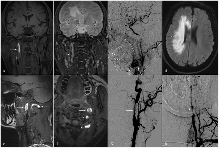
Results: Patients with mtCAD are significantly younger than sCAD with 120 cases of sCAD average aged 43.61 ± 12.75, while 28 cases of mtCAD average aged 35.68 ± 14.54. Patients with mtCAD had more cases of neck pain compared to sCAD. Patients with mtCAD had more cases of CAD at extracranial locations compared to sCAD. Patients with mtCAD had more cases of multiple site dissection compared to sCAD. Double lumen and intramural haematoma are the most common imaging findings with mtCAD patients having statistical significantly more cases of intramural haematoma and long tapering stenosis.
Conclusion: Patients with mtCAD were presented at a much younger age with symptoms of neck pain compared to sCAD. Patients with mtCAD predominantly presented at extracranial sites with more prominent features of multiple site dissection, intramural haematoma, and long tapering stenosis. These clinical and imaging features can translate into clinical practice guidelines for patients with CAD to improve the optimal functional outcome and reduce both morbidity and mortality.
Keywords: cervical manipulation; craniocervical arterial dissection; ischaemic stroke; minor trauma dissection; spontaneous dissections.
Copyright © 2022 Xu, Wu, Li, Xing, Chen, Chen, Tan, Zhou, Zhang and Zhang.
Conflict of interest statement
The authors declare that the research was conducted in the absence of any commercial or financial relationships that could be construed as a potential conflict of interest.
Figures
References
-
- Nagumo K, Nakamori A, Kojima S. [Spontaneous intracranial internal carotid artery dissection: 6 case reports and a review of 39 cases in the literature]. Rinsho Shinkeigaku. (2003) 43:313–21. - PubMed
LinkOut - more resources
Full Text Sources
Miscellaneous