. 2022 Mar 24:13:859929.
doi: 10.3389/fimmu.2022.859929. eCollection 2022.

Proteomics Analysis of Aqueous Humor and Rejected Graft in Pig-to-Non-Human Primate Corneal Xenotransplantation

Affiliations

Proteomics Analysis of Aqueous Humor and Rejected Graft in Pig-to-Non-Human Primate Corneal Xenotransplantation

Jae Won Oh et al. Front Immunol. .

Abstract

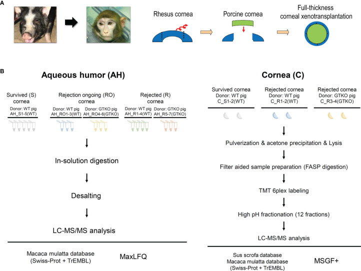
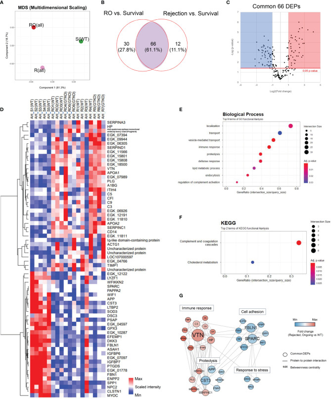
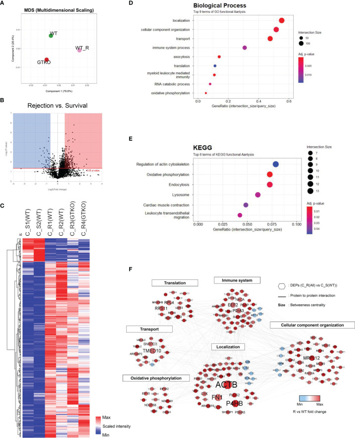
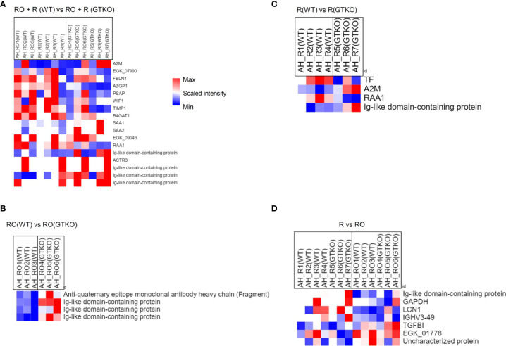
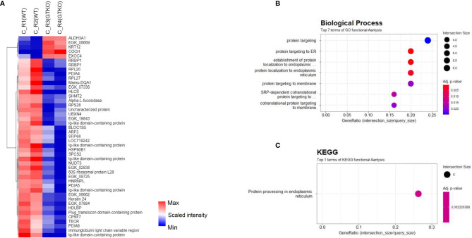
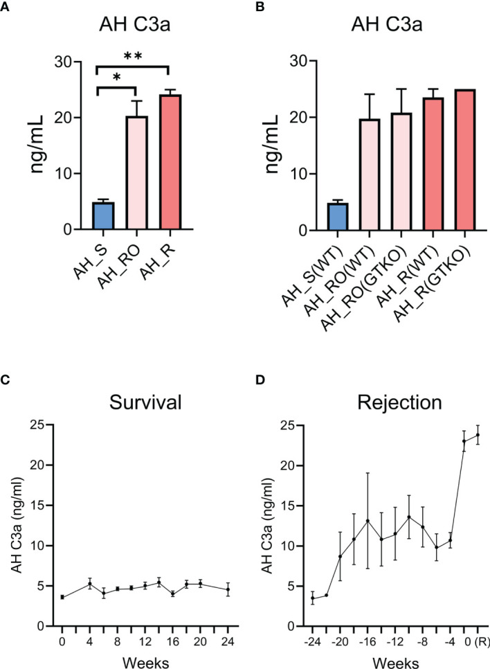
Although pig-to-non-human primate (NHP) corneal xenotransplantation has shown long-term graft survival, xenogeneic antigen-related immune responses are still stronger than allogeneic antigen-associated responses. Therefore, there is an unmet need to investigate major rejection pathways in corneal xenotransplantation, even with immunosuppression. This study aimed to identify biomarkers in aqueous humor for predicting rejection and to investigate rejection-related pathways in grafts from NHPs transplanted with porcine corneas following the administration of steroids combined with tacrolimus/rituximab. NHPs who had received corneas from wild-type (WT) or α-1,3-galactosyltransferase gene-knockout (GTKO) pigs were divided into groups with or without rejection according to clinical examinations. Liquid chromatography-mass spectrometry (LC-MS) was used to analyze the proteomes of corneal tissues or aqueous humor. The biological functions of differentially expressed proteins (DEPs) were assessed using Gene Ontology (GO) and Kyoto Encyclopedia of Genes and Genomes (KEGG) for pathways and protein-protein interaction network analysis. Among the 66 DEPs in aqueous humor, complement proteins (C3, C5, and C9) and cholesterol metabolic proteins (APOA1 and APOA2) were related to xenogeneic rejection as biomarkers, and alternative pathways of the complement system seemed to be important in xenogeneic graft rejection. Among the 416 DEPs of the cornea, NF-κB1 and proteosomes (PSMD7, PSMA5, and PSMD3) seemed to be related to xenogeneic graft rejection. Additionally, oxidative phosphorylation and leukocyte activation-related pathways are involved in rejection. Overall, our proteomic approach highlights the important role of NF-κB1, proteosomes, oxidative phosphorylation, and leukocyte activation-related inflammation in the cornea and the relevance of complement pathways of the aqueous humor as a predictive biomarker of xenogeneic rejection.

Keywords: aqueous humor; cornea; non-human primate (macaque); pig; proteomics; rejection; xenotransplantation.


Conflict of interest statement

The authors declare that the research was conducted in the absence of any commercial or financial relationships that could be construed as a potential conflict of interest.

Figures

Figure 1
Figure 1
(A) Schematic diagram depicting the procedure used for full-thickness corneal xenotransplantation. A clinically applicable porcine corneal donor button of 7.5 mm was used on the 7.0-mm non-human primate recipient bed in all cases. (B) Overall experimental workflow of the proteomics experiments. Aqueous humor (AH) and cornea were lysed and digested into peptides. Digested peptides were analyzed using LC-MS/MS. AH samples were subjected to LFQ analysis and cornea samples were subjected to TMT quantitative analysis. WT, wild type; GTKO, α-1,3-galactosyltransferase gene-knockout; LC, liquid chromatography; LFQ, label-free quantification; TMT, tandem mass tag.
Figure 2
Figure 2
Proteomic analysis of the aqueous humor (AH). Proteins from a non-human primate library (Macaca mulatta) with at least 2-fold significant (p < 0.05) changes in the rejection ongoing (RO) and rejection (R) groups [compared with the survival (S) group] were analyzed. (A) Principal component analysis (PCA) of AH protein expression in the R, RO, and S groups. (B) Venn diagram depicting the comparison of differentially expressed proteins (DEPs) in the RO and R groups compared with the S group. A set of 66 proteins (common DEPs) exhibited a significant and more than 2-fold change in both the RO and R groups (compared with the S group). (C) A volcano plot showing the differential level of proteins in the RO and R groups compared with the S group. (D) Cluster heat map of the 66 common DEPs identified in AH. (E, F) Gene Ontology biological process classification (E) and Kyoto Encyclopedia of Genes and Genomes (KEGG) pathway enrichment analyses (F) of the common DEPs. Node size represents the gene ratio; node color represents the adjusted p-value. (G) Network modeling of the common DEPs in AH. A network model showing the biological processes affected, including the immune response, proteolysis, cell adhesion, and response to stress. The colors of the nodes represent proteins whose levels were greatly increased (red) or decreased (blue) in the RO and R groups compared with the S group. Large nodes indicate a high degree of connectivity (betweenness centrality) with other proteins in the model. Connections between nodes (gray lines) indicate either regulatory roles or physical interactions between proteins. WT, wild type; GTKO, α-1,3-galactosyltransferase gene-knockout.
Figure 3
Figure 3
Proteomic analysis of the corneal tissues using a non-human primate (Macaca mulatta) library. Proteins with at least 1.5-fold significant (p < 0.05) changes in the rejection (R) groups [compared with the survival (S) group] were analyzed. (A) Principal component analysis of corneal tissue protein expression in the wild-type (WT) porcine cornea transplant group with rejection [C_R(WT)] or without rejection [survival; (C_S(WT)] and α-1,3‐galactosyltransferase gene‐knockout (GTKO) porcine cornea transplant group with rejection [C_R(GTKO)]. (B) A volcano plot showing the differential level of proteins in the R group compared with the S group. (C) Cluster heat map of DEPs identified in corneal tissue. (D, E) Gene Ontology biological process classification (D) and Kyoto Encyclopedia of Genes and Genomes (KEGG) pathway enrichment analyses (E) of the DEPs in corneal tissue. Node size represents the gene ratio; node color represents the adjusted p-value. (F) Network modeling of the DEPs in corneal tissue. The network model showing the biological processes affected, including the immune system, cellular component organization, and oxidative phosphorylation. The colors of the nodes represent proteins whose levels were greatly increased (red) or decreased (blue) in the R group compared with the S group. Large nodes indicate a high degree of connectivity (betweenness centrality) with other proteins in the model. Connections between nodes (gray lines) indicate either regulatory roles or physical interactions between proteins.
Figure 4
Figure 4
Proteomic analysis of the subgroups in aqueous humor (AH) using a non-human primate (Macaca mulatta) library. (A) Cluster heat map of differentially expressed proteins (DEPs) comparing subgroups transplanted with wild-type (WT) or α-1,3‐galactosyltransferase gene‐knockout (GTKO) porcine corneas when the rejection ongoing (RO) and rejection (R) groups were combined. (B) Cluster heat map of DEPs comparing subgroups transplanted with WT or GTKO porcine corneas in the RO group. (C) Cluster heat map of DEPs comparing subgroups transplanted with WT or GTKO porcine corneas in the R group. (D) Cluster heat map of DEPs comparing the R and RO groups regardless of donor corneal characteristics.
Figure 5
Figure 5
Proteomic analysis of the subgroups in corneal tissue. (A) Cluster heat map of differentially expressed proteins (DEPs) comparing subgroups transplanted with wild-type (WT) or α-1,3‐galactosyltransferase gene‐knockout (GTKO) porcine corneas in the rejection (R) group. (B, C) Gene Ontology biological process classification (B) and Kyoto Encyclopedia of Genes and Genomes (KEGG) pathway enrichment analyses (C) of the DEPs between subgroups transplanted with WT and GTKO porcine corneas in the R group. Node size represents the gene ratio; node color represents the adjusted p-value.
Figure 6
Figure 6
Validation of aqueous humor (AH) C3a as a putative biomarker for corneal xenograft rejection. (A) Concentration of aqueous humor (AH) C3a in the survival (AH_S), rejection ongoing (AH_RO), and rejection (AH_R) groups. The concentration of AH C3a was significantly higher in the rejection ongoing (RO) and rejection (R) groups compared with that in the survival (S) group (p = 0.034 and p = 0.002; Kruskal–Wallis test with Dunn’s multiple comparisons test). (B) Subgroup analysis of the concentration of AH C3a. AH_RO(WT) and AH_RO(GTKO) mean the AH of the RO subgroup transplanted with wild type (WT) and α-1,3‐galactosyltransferase gene‐knockout (GTKO) porcine cornea, respectively. AH_R(WT) and AH_R(GTKO) mean the AH of the R subgroup transplanted with WT and GTKO porcine cornea, respectively. The concentration did not differ according to the type of donor cornea (WT versus GTKO). (C) Time-dependent changes of AH C3a concentration in the survival group after xenotransplantation. The time point of xenotransplantation was set as the reference time point (0 week). (D) Time-dependent changes of AH C3a concentration in the rejection group after xenotransplantation. The time point at which xenograft rejection was set as the reference time point (0 week) and the AH C3a concentrations before rejection were shown. R, rejection. Data are presented as means ± standard error. *p < 0.05, **p < 0.01.

Similar articles

Cited by

References

    1. Tan DT, Dart JK, Holland EJ, Kinoshita S. Corneal Transplantation. Lancet (2012) 379(9827):1749–61. doi: 10.1016/s0140-6736(12)60437-1 - DOI - PubMed
    1. Gain P, Jullienne R, He Z, Aldossary M, Acquart S, Cognasse F, et al. . Global Survey of Corneal Transplantation and Eye Banking. JAMA Ophthalmol (2016) 134(2):167–73. doi: 10.1001/jamaophthalmol.2015.4776 - DOI - PubMed
    1. Yoon CH, Choi HJ, Kim MK. Corneal Xenotransplantation: Where Are We Standing? Prog Retin Eye Res (2021) 80:100876. doi: 10.1016/j.preteyeres.2020.100876 - DOI - PMC - PubMed
    1. Lai L, Kolber-Simonds D, Park KW, Cheong HT, Greenstein JL, Im GS, et al. . Production of Alpha-1,3-Galactosyltransferase Knockout Pigs by Nuclear Transfer Cloning. Science (2002) 295(5557):1089–92. doi: 10.1126/science.1068228 - DOI - PubMed
    1. Yoon CH, Choi SH, Choi HJ, Lee HJ, Kang HJ, Kim JM, et al. . Long-Term Survival of Full-Thickness Corneal Xenografts From A1,3-Galactosyltransferase Gene-Knockout Miniature Pigs in Non-Human Primates. Xenotransplantation (2020) 27(1):e12559. doi: 10.1111/xen.12559 - DOI - PubMed

Publication types