. 2022 Mar 24:13:791799.
doi: 10.3389/fimmu.2022.791799. eCollection 2022.

PgtE Enzyme of Salmonella enterica Shares the Similar Biological Roles to Plasminogen Activator (Pla) in Interacting With DEC-205 (CD205), and Enhancing Host Dissemination and Infectivity by Yersinia pestis

Affiliations

PgtE Enzyme of Salmonella enterica Shares the Similar Biological Roles to Plasminogen Activator (Pla) in Interacting With DEC-205 (CD205), and Enhancing Host Dissemination and Infectivity by Yersinia pestis

Qiao Li et al. Front Immunol. .

Abstract

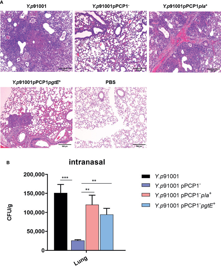
Yersinia pestis, the cause of plague, is a newly evolved Gram-negative bacterium. Through the acquisition of the plasminogen activator (Pla), Y. pestis gained the means to rapidly disseminate throughout its mammalian hosts. It was suggested that Y. pestis utilizes Pla to interact with the DEC-205 (CD205) receptor on antigen-presenting cells (APCs) to initiate host dissemination and infection. However, the evolutionary origin of Pla has not been fully elucidated. The PgtE enzyme of Salmonella enterica, involved in host dissemination, shows sequence similarity with the Y. pestis Pla. In this study, we demonstrated that both Escherichia coli K-12 and Y. pestis bacteria expressing the PgtE-protein were able to interact with primary alveolar macrophages and DEC-205-transfected CHO cells. The interaction between PgtE-expressing bacteria and DEC-205-expressing transfectants could be inhibited by the application of an anti-DEC-205 antibody. Moreover, PgtE-expressing Y. pestis partially re-gained the ability to promote host dissemination and infection. In conclusion, the DEC-205-PgtE interaction plays a role in promoting the dissemination and infection of Y. pestis, suggesting that Pla and the PgtE of S. enterica might share a common evolutionary origin.

Keywords: DEC-205 (CD205); PgtE; Salmonella enterica; Yersinia pestis; dissemination; evolution.


Conflict of interest statement

CP was employed by Genuv Inc. The remaining authors declare that the research was conducted in the absence of any commercial or financial relationships that could be construed as a potential conflict of interest.

Figures

Figure 1
Figure 1
PgtE in recombinant Y. pestis activates plasminogen to plasmin. The plasminogen activation activities of Y.p1419 pPCP1+, Y.p1419, Y.p1419 pgtE+ and E.coli, E.coli pla+ were compared. Y.p1418 was used as a positive control. PBS was used as negative control. The data presented were pooled from three independent experiments.
Figure 2
Figure 2
PgtE-expressing E. coli and Y. pestis invade alveolar macrophages and invade CHO-m-DEC-205. (A) PgtE-expressing E. coli were examined for their ability to enter alveolar macrophages. The bacteria used were E. coli, E. coli pla+ and E. coli pgtE+. (B) PgtE-expressing Y. pestis were examined for their ability to enter alveolar macrophages. The bacteria used were Y.p1418, Y.p1419, Y.p1419 pla+, Y.p1419 pgtE+ .The number of phagocytized bacteria was determined by evaluating the CFUs on the plates after two days. The data presented were collective from three independent experiments. *P < 0.05, **P < 0.01, ***P < 0.001. (C) PgtE-expressing E. coli invade the CHO cell line expressing CD205. Epithelial CHO cells expressing or not expressing CD205 (CHO and CD205, respectively) were infected with PgtE- and Pla-expressing E. coli. Y. pseudotuberculosis (Y1); E. coli, E. coli pla+ , and E. coli pgtE+ were examined for their abilities to invade CHO/CHO-m-DEC-205 cells during a gentamicin protection assay, in presence or absence of anti-DEC-205 (5 μg/ml). The numbers of phagocytosed bacteria were determined by counting the bacterial CFUs on the plates the next day. The data presented were pooled from three independent experiments. *P < 0.05, **P < 0.01, ***P < 0.001. (D) PgtE-expressing Y. pestis invades the CHO cell line expressing CD205. Epithelial CHO cells expressing or not expressing CD205 (CHO and CD205, respectively) were infected with PgtE- and Pla-expressing Y. pestis. Y. pseudotuberculosis (Y1), Y.p1419, Y.p1419 pla+ and Y.p1419 pgtE+ were examined for their abilities to invade CHO/CHO-m-DEC-205 cells during a gentamicin protection assay, in presence or absence of anti-DEC-205 (5 μg/ml). The numbers of phagocytosed bacteria were determined by counting the bacterial CFUs on the plates after two days. The data presented were pooled from three independent experiments. *P < 0.05, **P < 0.01, ***P < 0.001.
Figure 3
Figure 3
PgtE expressed in Y. pestis enhances the ability to promote host dissemination. Y.p91001, Y.p91001pPCP1-, Y.p91001 pPCP1-pla+ and Y.p91001 pPCP1-pgtE+ were used to challenge mice via the intranasal route. After 72 hours of infection, the liver and spleen were collected and homogenized. The bacterial loads were quantified by counting the bacteria colonies on the plates after two days. (The data shown were obtained from three independent experiments. *P < 0.05, **P < 0.01.
Figure 4
Figure 4
Mice infected intranasally with PgtE-expressing Y. pestis are more susceptible to death compared with pPCP1 plasmid cured and pla-deleted Y. pestis. (A) Y.p91001, Y.p91001pPCP1-, Y.p91001 pPCP1-pla+ and Y.p91001 pPCP1-pgtE+ were used to challenge mice via the intranasal route. The mice were monitored for 12 days, and the log-rank test was performed. (B) Y.p91001, Y.p91001pla-, Y.p91001pla- +pla+ , Y.p91001pla- +pgtE+ were used to challenge mice via the intranasal route. The data shown were obtained from three independent experiments.
Figure 5
Figure 5
The expression of PgtE in Y. pestis was able to enhance the inflammatory lesions in the lungs from C57BL/6 mice. (A)Y.p91001, Y.p91001pPCP1-, Y.p91001 pPCP1-pla+ and Y.p91001 pPCP1-pgtE+ were used to challenge mice via the intranasal route. Lung damage was examined by hematoxylin and eosin (H & E) staining of formalin-fixed sections 48 hours after infection. C57BL/6 mice were inoculated with PBS (mock), Y. pestis Y.p91001, Y.p91001pPCP1-, Y.p91001 pPCP1-pla+ and Y.p 91001pPCP1-pgtE+ strains Representative images of inflammatory lesions are shown. (B) The bacteria amount in the lung tissues of the mice infected by Y.p91001, Y.p91001pPCP1-, Y.p91001 pPCP1-pla+ and Y.p91001 pPCP1-pgtE+ were examined 8 hours after infection. **P < 0.01, ***P < 0.001.

Similar articles

References

    1. Perry RD, Fetherston JD. Yersinia Pestis–Etiologic Agent of Plague. Clin Microbiol Rev (1997) 10:35–66. doi: 10.1128/CMR.10.1.35 - DOI - PMC - PubMed
    1. Prentice Michael B, Tom G, Cooper A. Was the Black Death Caused by Yersinia Pestis ? Lancet Infect Dis (2004) 4:72. doi: 10.1016/s1473-3099(04)00923-5 - DOI - PubMed
    1. Bos Kirsten I, Schuenemann Verena J, Golding GB, Burbano Hernán A, Waglechner N, Coombes Brian K, et al. . A Draft Genome of Yersinia Pestis From Victims of the Black Death. Nature (2011) 478:506–10. doi: 10.1038/nature10549 - DOI - PMC - PubMed
    1. Spyrou MA, Tukhbatova RI, Feldman M, Drath J, Kacki S, Beltran de Heredia J, et al. . Historical Y. Pestis Genomes Reveal the European Black Death as the Source of Ancient and Modern Plague Pandemics. Cell Host Microbe (2016) 19:874–81. doi: 10.1016/j.chom.2016.05.012 - DOI - PubMed
    1. Sebbane F, Uversky VN, Anisimov AP. Yersinia Pestis Plasminogen Activator. Biomolecules (2020) 10:1554. doi: 10.3390/biom10111554 - DOI - PMC - PubMed