. 2022 Mar 23:13:817040.
doi: 10.3389/fimmu.2022.817040. eCollection 2022.

Enhanced Migratory Ability of Neutrophils Toward Epidermis Contributes to the Development of Psoriasis via Crosstalk With Keratinocytes by Releasing IL-17A

Affiliations

Enhanced Migratory Ability of Neutrophils Toward Epidermis Contributes to the Development of Psoriasis via Crosstalk With Keratinocytes by Releasing IL-17A

Xiu-Ting Liu et al. Front Immunol. .

Abstract

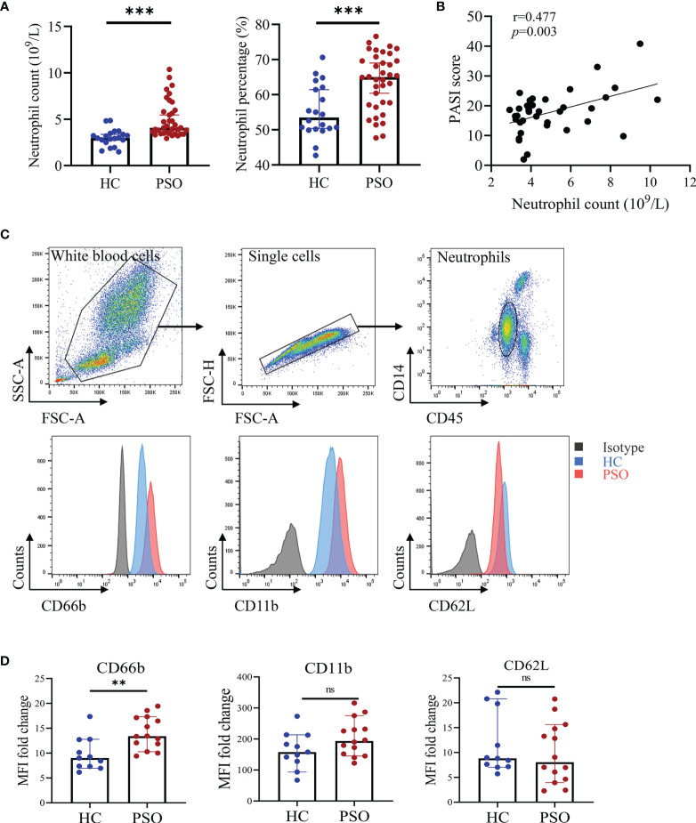
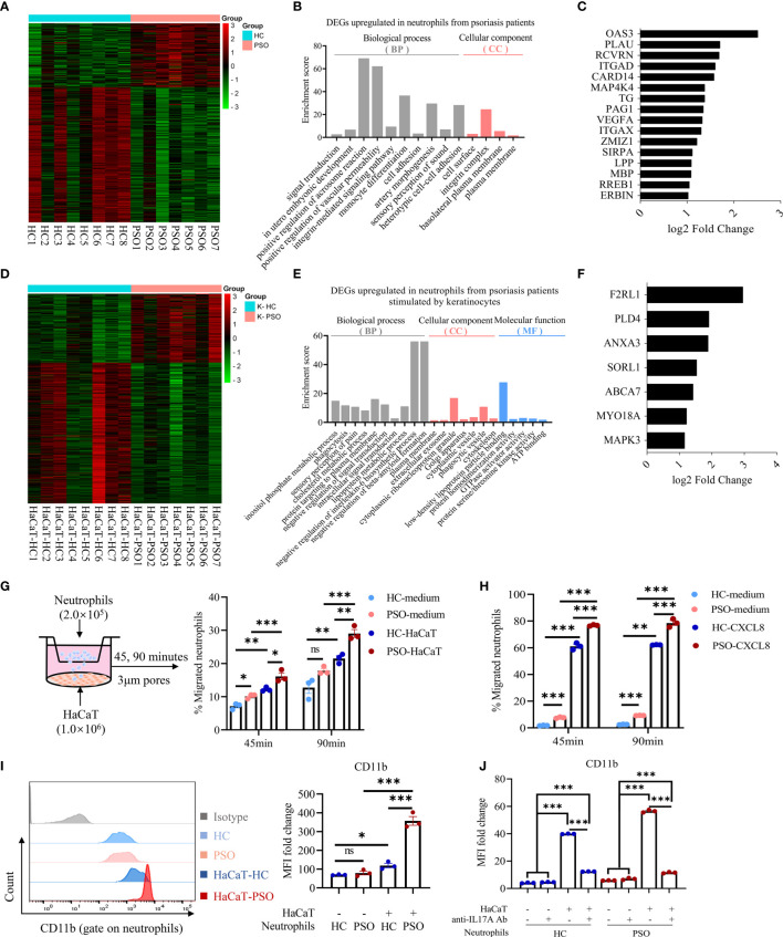
Microabscess of neutrophils in epidermis is one of the histological hallmarks of psoriasis. The axis of neutrophil-keratinocyte has been thought to play a critical role in the pathogenesis of psoriasis. However, the features and mechanism of interaction between the two cell types remain largely unknown. Herein, we found that blood neutrophils were increased in psoriasis patients, positively correlated with disease severity and highly expressed CD66b, but not CD11b and CD62L compared to healthy controls. Keratinocytes expressed high levels of psoriasis-related inflammatory mediators by direct and indirect interaction with neutrophils isolated from psoriasis patients and healthy controls. The capacity of neutrophils in provoking keratinocytes inflammatory response was comparable between the two groups and is dependent on IL-17A produced by itself. Neutrophils isolated from psoriasis patients displayed more transcriptome changes related to integrin and increased migration capacity toward keratinocytes with high CD11b expression on cell surface. Of interest, neutrophils were more susceptible to keratinocyte stimulation than to fibroblasts and human umbilical vein endothelial cells (HUVECs) in terms of CD11b expression and the production of ROS and NETs. In conclusion, neutrophils from psoriasis patients gain a strong capacity of IL-17A production and integrins expression that possibly facilitates their abilities to promote production of psoriasis-related inflammatory mediators and migration, a phenomenon likely induced by their interaction with keratinocytes but not with fibroblasts. These findings provide a proof-of-concept that development of new drugs targeting migration of neutrophils could be a more specific and safe solution to treat psoriasis.

Keywords: interleukin-17A; keratinocytes; migration; neutrophils; psoriasis.


Conflict of interest statement

The authors declare that the research was conducted in the absence of any commercial or financial relationships that could be construed as a potential conflict of interest.

Figures

Figure 1
Figure 1
Blood neutrophils are increased and pre-activated in psoriasis patients. (A) Absolute numbers and percentages of neutrophils (in white blood cells) in peripheral blood of healthy controls (HC, n = 20) and psoriasis patients (PSO, n = 36). (B) The correlation of the absolute numbers of blood neutrophils and PASI score in psoriasis patients (n = 36). (C) Representative flow plots showing gating strategy of blood neutrophils and CD66b, CD11b, and CD62L expressions in stained HC (blue) and PSO (red) samples versus isotype control (gray). (D) Fold changes of MFI (versus isotype) of CD66b, CD11b, and CD62L on neutrophils of HC (n = 11) and PSO (n = 14). Data are presented as median ± 95% CI. **p < 0.01; ***p < 0.001, by Mann–Whitney test. The correlation analysis was performed by Spearman correlation coefficient. CI, confidence interval; HC, healthy controls; MFI, median fluorescence intensity; ns, not significant; PASI, psoriasis area and severity index; PSO, psoriasis.
Figure 2
Figure 2
Neutrophils promote inflammatory mediators expression by keratinocytes and cell apoptosis. (A) A scheme of direct coculture system (left panel). The mRNA expression of psoriasis-related inflammatory mediators in HaCaT 24 h after direct coculture with neutrophil (right panel). (B) Protein levels of CXCL8 in the supernatants determined by ELISA 8 h after co-culture. (C) A scheme of indirect coculture system using transwell inserts (left panel). The mRNA expression of psoriasis related-inflammatory mediators (C, right panel) and the protein levels of CXCL8 in the supernatants (D). (E) Apoptosis rate of HaCaT cells cultured with neutrophils in indirect contact system for 24 h. CTR (control) refers to HaCaT cells without neutrophils. The p-values depicted as asterisk in (A, C) refers to comparisons with control. Data are representative of three or more independent experiments. Data are presented as mean ± SEM. *p < 0.05; **p < 0.01; ***p < 0.001, by one-way ANOVA. ELISA, enzyme-linked immunosorbent assay; ns, not significant.
Figure 3
Figure 3
Neutrophils promote the inflammatory response of keratinocytes through release of IL-17A. (A) Representative image of IL-17A expression in blood neutrophils examined by western blot (left panel) and quantification of western blot analysis from healthy controls (HC, n = 20) and psoriasis patients (PSO, n = 25) (right panel). (B) mRNA expression of IL-17A in blood neutrophils from HC (n = 21) and PSO (n = 22). (C) Representative immunofluorescence images of MPO (green),IL-17A (red) and DAPI (blue) expression in blood neutrophils from HC (n = 6) or PSO (n = 5), scale bar = 20 µm. Quantification analysis of IL-17A expression in neutrophils from HC and PSO. (D) Protein levels of IL-17A in the supernatant determined by ELISA at 8 h from coculture system of HaCaT alone, HaCaT plus healthy neutrophils or HaCaT plus psoriatic neutrophils. (E) mRNA expression of IL-17A in HaCaT cells stimulated by neutrophils (square) and in neutrophils (circle) from psoriasis patients. (F) mRNA expression of inflammatory mediators in HaCaT 8 h after directly coculture with neutrophils with and without anti-IL-17A Abs (12.5 μg/ml). Data are representative of three or more independent experiments. Data are presented mean ± SEM and analyzed by the two-tailed Student’s t-test or one-way ANOVA. *p < 0.05; **p < 0.01; ***p < 0.001; Abs, antibodies; ns, not significant.
Figure 4
Figure 4
Neutrophils gain migration phenotype and capacity after coculture with keratinocytes. (A) Heatmap showing DEGs of RNA-Seq results in PSO (n = 7) versus HC (n = 8) neutrophils cultured alone for 8 h. (B) GO category and (C) migration-related DEGs upregulated in PSO neutrophils. (D) Heatmap showing DEGs in PSO versus HC neutrophils after indirect coculture with HaCaT for 8 h. (E) GO category and (F) phagocytosis-related DEGs upregulated in PSO neutrophils. (G) A scheme of transwell assay. Peripheral blood neutrophils (2 × 10⁵ cells) in the upper chamber were allowed to migrate through 3 μm pores of transwell membrane towards the lower chamber with complete medium or pre-seeded with HaCaT, and the rates of neutrophils migration at indicated time points were analyzed by flow cytometry. HC-medium, HC neutrophils with medium, PSO-medium, PSO neutrophils with medium, HC-HaCaT, HC neutrophils with HaCaT, PSO neutrophils with HaCaT. (H) Transwell assay comparing the migration of HC or PSO neutrophils towards complete medium with or without CXCL8 protein (200 ng/ml). (I) Representative images of flow cytometry and quantification of MFI fold changes of CD11b expression on neutrophils cultured with or without HaCaT in an indirect manner for 24 h. (J) Quantification of MFI fold changes of CD11b expression on neutrophils cultured as described in (I) with or withoutanti-IL-17A Abs (12.5 μg/ml). Data are presented as mean ± SEM, one-way ANOVA. *p < 0.05; **p < 0.01; ***p < 0.001. DEGs, differently expressed genes; GO, Gene Ontology; MFI, median fluorescence intensity; ns, not significant; RNA-Seq, RNA-sequencing.
Figure 5
Figure 5
Neutrophils are particularly activated by keratinocytes. (A, B) Representative graph and quantification of CD11b expression (A) and ROS (B) in neutrophils stimulated by HaCaT, fibroblast and HUVEC. (C) Representative immunofluorescence staining for MPO (green), DAPI (blue) and merged images of neutrophils stimulated by vehicle, PMA (100 nmol/ml, positive control), HaCaT, fibroblasts and HUVECs, respectively. Scales bar = 20 µm. White arrow indicates NETs. Data are representative of three or more independent experiments. Data are presented as mean ± SEM, analyzed by one-way ANOVA. *p < 0.05; **p < 0.01; ***p < 0.001. HUVEC, human umbilical vein endothelial cells; NETs, neutrophil extracellular traps; ROS, reactive oxygen species.

Similar articles

Cited by

References

    1. Metzemaekers M, Gouwy M, Proost P. Neutrophil Chemoattractant Receptors in Health and Disease: Double-Edged Swords. Cell Mol Immunol (2020) 17(5):433–50. doi: 10.1038/s41423-020-0412-0 - DOI - PMC - PubMed
    1. Chiang CC, Cheng WJ, Korinek M, Lin CY, Hwang TL. Neutrophils in Psoriasis. Front Immunol (2019) 10:2376. doi: 10.3389/fimmu.2019.02376 - DOI - PMC - PubMed
    1. Wolk K, Brembach TC, Simaite D, Bartnik E, Cucinotta S, Pokrywka A, et al. . Activity and Components of the Granulocyte Colony-Stimulating Factor Pathway in Hidradenitis Suppurativa. Br J Dermatol (2021) 185(1):164–76. doi: 10.1111/bjd.19795 - DOI - PubMed
    1. Shi Z, Garcia-Melchor E, Wu X, Yu S, Nguyen M, Rowland DJ, et al. . Differential Requirement for CCR6 in IL-23-Mediated Skin and Joint Inflammation. J Invest Dermatol (2020) 140(12):2386–97. doi: 10.1016/j.jid.2020.03.965 - DOI - PubMed
    1. Sumida H, Yanagida K, Kita Y, Abe J, Matsushima K, Nakamura M, et al. . Interplay Between CXCR2 and BLT1 Facilitates Neutrophil Infiltration and Resultant Keratinocyte Activation in a Murine Model of Imiquimod-Induced Psoriasis. J Immunol (2014) 192(9):4361–9. doi: 10.4049/jimmunol.1302959 - DOI - PubMed