Review
. 2022 Mar 23:13:868679.
doi: 10.3389/fimmu.2022.868679. eCollection 2022.

Cardiovascular Dysfunction in COVID-19: Association Between Endothelial Cell Injury and Lactate

Affiliations
Review

Cardiovascular Dysfunction in COVID-19: Association Between Endothelial Cell Injury and Lactate

Kun Yang et al. Front Immunol. .

Abstract

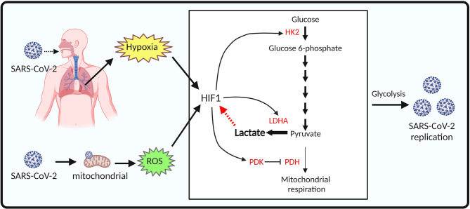
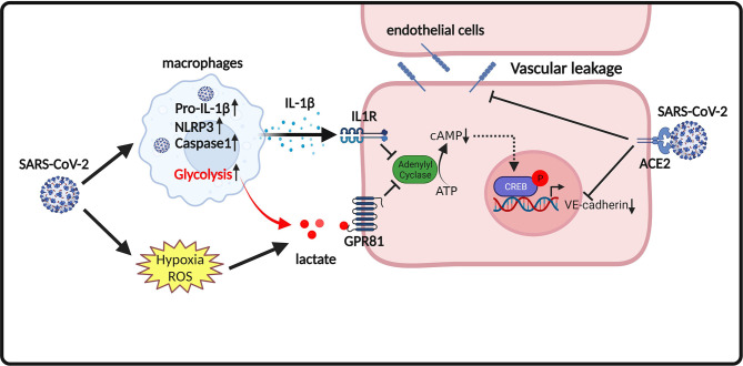
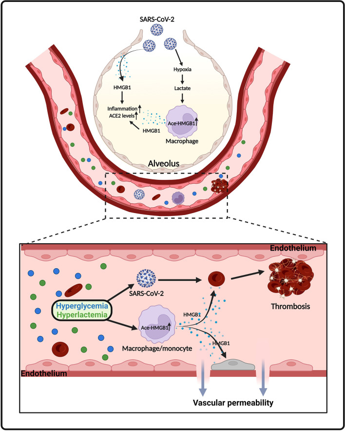
Coronavirus disease 2019 (COVID-19), an infectious respiratory disease propagated by a new virus known as Severe Acute Respiratory Syndrome Coronavirus-2 (SARS-CoV-2), has resulted in global healthcare crises. Emerging evidence from patients with COVID-19 suggests that endothelial cell damage plays a central role in COVID-19 pathogenesis and could be a major contributor to the severity and mortality of COVID-19. Like other infectious diseases, the pathogenesis of COVID-19 is closely associated with metabolic processes. Lactate, a potential biomarker in COVID-19, has recently been shown to mediate endothelial barrier dysfunction. In this review, we provide an overview of cardiovascular injuries and metabolic alterations caused by SARS-CoV-2 infection. We also propose that lactate plays a potential role in COVID-19-driven endothelial cell injury.

Keywords: COVID-19; HMGB1 (High mobility group box 1); aerobic glycolytic metabolism; cardiovascular dysfunction; endothelial cell; lactate; thrombosis; vascular permeability.


Conflict of interest statement

The authors declare that the research was conducted in the absence of any commercial or financial relationships that could be construed as a potential conflict of interest.

Figures

Figure 1
Figure 1
Proposed model of aerobic glycolysis activation in SARS-CoV-2 infected endothelial cells. SARS-CoV-2 infection of pulmonary tissues leads to hypoxia. SARS-CoV-2 infection also causes mitochondrial dysfunction and reactive oxygen species (ROS) production in endothelial cells. Both hypoxia and ROS mediate HIF-1 stabilization. Enzymes involved in glycolysis, including hexokinase (HK), pyruvate kinase 2 (PKM2), lactate dehydrogenase (LDH) are upregulated by HIF-1 signaling, resulting in increased lactate production and SARS-CoV-2 replication in endothelial cells.
Figure 2
Figure 2
Proposed model of endothelium permeability induced by lactate/GPR81 signaling and SARS-CoV-2 infection. SARS-CoV-2 infection promotes the release of the pro-inflammatory cytokine IL-1β. IL-1β suppresses cAMP formation and CREB-mediated transcription of VE-cadherin in endothelial cells. SARS-CoV-2 infection also increases lactate production. Lactate activates GPR81 and reduces cAMP generation and CREB-mediated transcription of VE-cadherin in endothelial cells. In addition, SARS-CoV-2 spike proteins directly disorganize VE-cadherin complex and suppress VE-cadherin transcription in endothelial cells. Disruption of VE-cadherin complex is responsible for vascular permeability in COVID-19.
Figure 3
Figure 3
Proposed model of platelet activation, thrombosis and endothelial cell injury induced by SARS-CoV-2 infection and lactate. Binding of SARS-CoV-2 spike protein to ACE2 leads to MAPK signaling activation and subsequent platelet activation. Activated platelets release coagulation factors and cytokines to promote thrombosis. Internalization of SARS-CoV-2 virions induces the release of extracellular vesicles from platelets to facilitate thrombosis. In addition, lactate (acidity) also contributes to thrombosis by promoting activation of platelets, endothelial cells, and NETs.
Figure 4
Figure 4
Proposed model of HMGB1 release in SARS-CoV-2 infection. SARS-CoV-2 infection causes death of epithelial cells and release of HMGB1. Lactate, derived from aerobic glycolysis, also promotes HMGB1 acetylation and release from macrophages/monocytes. Elevated levels of HMGB1 further promotes inflammatory responses, ACE2 expression, endothelium permeability and thrombosis in COVID-19.

Similar articles

  • Physical interventions to interrupt or reduce the spread of respiratory viruses.
    Jefferson T, Dooley L, Ferroni E, Al-Ansary LA, van Driel ML, Bawazeer GA, Jones MA, Hoffmann TC, Clark J, Beller EM, Glasziou PP, Conly JM. Jefferson T, et al. Cochrane Database Syst Rev. 2023 Jan 30;1(1):CD006207. doi: 10.1002/14651858.CD006207.pub6. Cochrane Database Syst Rev. 2023. PMID: 36715243 Free PMC article.
  • Systemic Inflammatory Response Syndrome.
    Baddam S, Burns B. Baddam S, et al. 2025 Jun 20. In: StatPearls [Internet]. Treasure Island (FL): StatPearls Publishing; 2025 Jan–. 2025 Jun 20. In: StatPearls [Internet]. Treasure Island (FL): StatPearls Publishing; 2025 Jan–. PMID: 31613449 Free Books & Documents.
  • Antibody tests for identification of current and past infection with SARS-CoV-2.
    Fox T, Geppert J, Dinnes J, Scandrett K, Bigio J, Sulis G, Hettiarachchi D, Mathangasinghe Y, Weeratunga P, Wickramasinghe D, Bergman H, Buckley BS, Probyn K, Sguassero Y, Davenport C, Cunningham J, Dittrich S, Emperador D, Hooft L, Leeflang MM, McInnes MD, Spijker R, Struyf T, Van den Bruel A, Verbakel JY, Takwoingi Y, Taylor-Phillips S, Deeks JJ; Cochrane COVID-19 Diagnostic Test Accuracy Group. Fox T, et al. Cochrane Database Syst Rev. 2022 Nov 17;11(11):CD013652. doi: 10.1002/14651858.CD013652.pub2. Cochrane Database Syst Rev. 2022. PMID: 36394900 Free PMC article.
  • Rapid, point-of-care antigen tests for diagnosis of SARS-CoV-2 infection.
    Dinnes J, Sharma P, Berhane S, van Wyk SS, Nyaaba N, Domen J, Taylor M, Cunningham J, Davenport C, Dittrich S, Emperador D, Hooft L, Leeflang MM, McInnes MD, Spijker R, Verbakel JY, Takwoingi Y, Taylor-Phillips S, Van den Bruel A, Deeks JJ; Cochrane COVID-19 Diagnostic Test Accuracy Group. Dinnes J, et al. Cochrane Database Syst Rev. 2022 Jul 22;7(7):CD013705. doi: 10.1002/14651858.CD013705.pub3. Cochrane Database Syst Rev. 2022. PMID: 35866452 Free PMC article.
  • Early spontaneous breathing for acute respiratory distress syndrome in individuals with COVID-19.
    Hohmann F, Wedekind L, Grundeis F, Dickel S, Frank J, Golinski M, Griesel M, Grimm C, Herchenhahn C, Kramer A, Metzendorf MI, Moerer O, Olbrich N, Thieme V, Vieler A, Fichtner F, Burns J, Laudi S. Hohmann F, et al. Cochrane Database Syst Rev. 2022 Jun 29;6(6):CD015077. doi: 10.1002/14651858.CD015077. Cochrane Database Syst Rev. 2022. PMID: 35767435 Free PMC article.

Cited by

References

    1. Gavriatopoulou M, Ntanasis-Stathopoulos I, Korompoki E, Fotiou D, Migkou M, Tzanninis IG, et al. . Emerging Treatment Strategies for COVID-19 Infection. Clin Exp Med (2021) 21(2):167–79. doi: 10.1007/s10238-020-00671-y - DOI - PMC - PubMed
    1. Huang C, Wang Y, Li X, Ren L, Zhao J, Hu Y, et al. . Clinical Features of Patients Infected With 2019 Novel Coronavirus in Wuhan, China. Lancet (2020) 395(10223):497–506. doi: 10.1016/S0140-6736(20)30183-5 - DOI - PMC - PubMed
    1. Terpos E, Ntanasis-Stathopoulos I, Elalamy I, Kastritis E, Sergentanis TN, Politou M, et al. . Hematological Findings and Complications of COVID-19. Am J Hematol (2020) 95(7):834–47. doi: 10.1002/ajh.25829 - DOI - PMC - PubMed
    1. Mohamed K, Rzymski P, Islam MS, Makuku R, Mushtaq A, Khan A, et al. . COVID-19 Vaccinations: The Unknowns, Challenges, and Hopes. J Med Virol (2022) 94(4):1336–49. doi: 10.1002/jmv.27487 - DOI - PMC - PubMed
    1. Dan S, Pant M, Upadhyay SK. The Case Fatality Rate in COVID-19 Patients With Cardiovascular Disease: Global Health Challenge and Paradigm in the Current Pandemic. Curr Pharmacol Rep (2020) p:1–10. doi: 10.1007/s40495-020-00239-0 - DOI - PMC - PubMed

Publication types