. 2022 Mar 23:13:816955.
doi: 10.3389/fgene.2022.816955. eCollection 2022.

The Ovarian Transcriptome at the Early Stage of Testis Removal-Induced Male-To-Female Sex Change in the Protandrous Black Porgy Acanthopagrus schlegelii

Affiliations

The Ovarian Transcriptome at the Early Stage of Testis Removal-Induced Male-To-Female Sex Change in the Protandrous Black Porgy Acanthopagrus schlegelii

Peng-Wei Tseng et al. Front Genet. .

Abstract

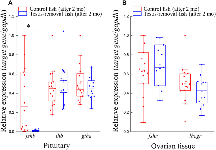
Unlike gonochoristic fishes, sex is fixed after gonadal differentiation (primary sex determination), and sex can be altered in adults (secondary sex determination) of hermaphroditic fish species. The secondary sex determination of hermaphroditic fish has focused on the differences between testicular tissue and ovarian tissue during the sex change process. However, comprehensive studies analyzing ovarian tissue or testicular tissue independently have not been performed. Hermaphroditic black porgy shows a digonic gonad (ovarian tissue with testicular tissue separated by connective tissue). Protandrous black porgy has stable maleness during the first two reproductive cycles (<2 years old), and approximately 50% enter femaleness (natural sex change) during the third reproductive cycle. Precocious femaleness is rarely observed in the estradiol-17β (E2)-induced female phase (oocytes maintained at the primary oocyte stage), and a reversible female-to-male sex change is found after E2 is withdrawn in <2-year-old fish. However, precocious femaleness (oocytes entering the vitellogenic oocyte stage) is observed in testis-removed fish in <2-year-old fish. We used this characteristic to study secondary sex determination (femaleness) in ovarian tissue via transcriptomic analysis. Cell proliferation analysis showed that BrdU (5-bromo-2'-deoxyuridine)-incorporated germline cells were significantly increased in the testis-removed fish (female) compared to the control (sham) fish (male) during the nonspawning season (2 months after surgery). qPCR analysis showed that there were no differences in pituitary-releasing hormones (lhb and gtha) in pituitary and ovarian steroidogenesis-related factors (star, cyp11a1, hsd3b1, and cyp19a1a) or female-related genes (wnt4a, bmp15, gdf9, figla, and foxl2) in ovarian tissues between intact and testis-removed fish (2 months after surgery). Low expression of pituitary fshb and ovarian cyp17a1 was found after 2 months of surgery. However, we did find small numbers of genes (289 genes) showing sexual fate dimorphic expression in both groups by transcriptomic analysis (1 month after surgery). The expression profiles of these differentially expressed genes were further examined by qPCR. Our present work identified several candidate genes in ovarian tissue that may be involved in the early period of secondary sex determination (femaleness) in black porgy. The data confirmed our previous suggestion that testicular tissue plays an important role in secondary sex determination in protandrous black porgy.

Keywords: estrogen; hermaphroditic fish; ovotestis; sex change; sex determination; sex differentiation; sexual fate.


Conflict of interest statement

The authors declare that the research was conducted in the absence of any commercial or financial relationships that could be construed as a potential conflict of interest.The authors declare that the research was conducted in the absence of any commercial or financial relationships that could be construed as a potential conflict of interest.

Figures

FIGURE 1
FIGURE 1
Schematic diagram of sex determination and gonadal differentiation in teleosts. Sex determination is a process to influence sexual fate. In gonochoristic fish, sex determination resulted in undifferentiated gonad to differentiated gonad (testis or ovary). Then the testis or ovary develops into a mature phase (functional sex). In syngonic fish, primary sex determination resulted in undifferentiated gonad to differentiated gonad, then the ovary develops into a mature phase (functional primary sex). In the natural sex change process, secondary sex determination caused primary sex to secondary sex (male-to-female or female-to-male). In digonic fish, such as black porgy, primary sex determination resulted in the change of undifferentiated gonad to differentiated gonad (a bisexual gonad, testis with an ovary), then the gonad develops into functional primary sex (mature testis with regressed ovary). In the natural sex change process, secondary sex determination caused primary sex to secondary sex. Undiff. gonad, undifferentiated gonad; Diff. gonad, differentiated gonad.
FIGURE 2
FIGURE 2
The morphology and histology analysis during testis removal-induced sex change in black porgy. In 1-year-old fish, testis excision was performed during the non-spawning season (Aug). We collected gonad during the pre-spawning season (2 months after surgery, Oct) for cell proliferation/histology (F–S) and second spawning season (Jan) for morphological analysis (A–E). For cell proliferation assay, BrdU (30 mg/kg BW) was injected on Day 7 and Day 3 before gonadal tissue was collected (2 months after surgery, Oct). Sertoli cell-expressing Dmrt1 was used to identify the testis-removed fish has a remnant testicular tissue or not. The gonad of control fish (A) and testis-removed fish (B) were collected at 2 months after surgery. The dash line (B) represents the branch of the pair gonad. The gonad of control fish (C) and testis-removed fish (D) were collected during the second spawning season. (E) The testis-removed fish has a remnant testicular tissue during the second spawning season. (F–I) H&E staining of the gonad that was collected at 2 months after surgery from the control fish and testis-removed fish. (G) and (I) are the high-magnification images of (F) and (H), respectively. (J–M) Gonadal IHC staining of Dmrt1 in the control fish and testis-removed fish (2 months after surgery). (K) and (M) are the high-magnification images of (J) and (L), respectively. (N–R) Gonadal IHC staining of BrdU in the control fish and testis-removed fish (2 months after surgery). (O,R) and (Q) are the high-magnification images of (N) and (P), respectively. (S) The results indicate that the oogonia have significantly increased proliferating activity in the testis-removed fish compared with the control fish. TT, testicular tissue; OT, ovarian tissue; CT, connected tissue; OG, oogonia; PO, primary oocyte, EGC, early germ cell; SG, spermatogonia; Sc, Sertoli cell. Asterisks indicate significant differences by Student’s t-test (***, p < 0.001).
FIGURE 3
FIGURE 3
Genes expression profiles of gths (fshb, lhb, and gtha) and gthrs (fshr and lhcgr) after testis-removal induced femaleness. There was no difference in cell composition of ovarian tissue in the control fish and testis-removed fish at 2 months after surgery. To understand the change of gths and gthrs in secondary sex determination, qPCR analysis for pituitary and ovarian tissue was used, respectively. Relative expression was normalized with gapdh, and the highest sample value of each gene in the control fish was defined as 1. Asterisks indicate significant differences by Student’s t-test (*, p < 0.05).
FIGURE 4
FIGURE 4
Expression profiles of ovarian steroidogenesis-related factors and female-related genes after testis-removal induced femaleness. To clarify the effect of sex steroids and female-related genes in secondary sex determination, we collected the ovarian tissue 2 months after surgery. The relative expression profiles of (A) ovarian steroidogenesis-related genes (star, cyp11a1, hsd3b1, cyp17a1, and cyp19a1a) and (B) potential female-related genes (wnt4a, bmp15, gdf9, figla, and foxl2) were detected using qPCR. Relative expression was normalized with gapdh, and the highest sample value of each gene in the control fish was defined as 1. Asterisks indicate significant differences by Student’s t-test (***, p < 0.001).
FIGURE 5
FIGURE 5
Volcano plot of differentially expressed genes in control fish and testis-removed fish. To find the genetic information during secondary sex determination, we performed differential expression analysis in the control fish and testis-removed fish with DESeq2. Genes with “adjusted p-value < 0.05” and “|log2Fold Change| > 1” were considered the DEGs (differentially expressed genes). The up-regulated (101 genes) and down-regulated (188 genes) DEGs in testis-removed fish were shown in red and blue dots, respectively. The gray dots represented the genes not belonging to DEGs.
FIGURE 6
FIGURE 6
KEGG pathway classification of DEGs. The x-axis indicates the number of DEGs (differentially expressed genes) matched in KEGG (Kyoto Encyclopedia of Genes and Genomes) pathway. The y-axis indicates the 33 KEGG subcategories.
FIGURE 7
FIGURE 7
Functional annotation of DEGs in the GO database. The counts of DEGs (differentially expressed genes) matched in level 2 GO (Gene Ontology) categories. The x-axis indicates 29 levels 2 GO categories. The y-axis indicates the number of DEGs that were matched.
FIGURE 8
FIGURE 8
Validation of transcriptomic data by qPCR. The 14 DEGs (differentially expressed genes) were selected randomly to detect the expression profiles. Some genes were not matched in any databases, and we named these genes DN1472, DN5641, DN16070, DN597, DN5503, and DN13274. The qPCR results were consistent with the transcriptomic data. y-Axis indicates Log2 (Gene expression profile of testis-removed fish/Gene expression profile of control fish).
FIGURE 9
FIGURE 9
Summary of sex-related genes involved in sex determination, differentiation, and development in black porgy. Primary sex determination and gonadal differentiation occurred around 3–4 months after hatching. But the exact timing and genes of primary sex determination are still not clear. The fish has functional males during the first two reproductive cycles. After secondary sex determination, we can observe developing ovaries with rudimentary testis. However, the key factors that activate secondary sex determination are still not clear. DEGs (differentially expressed genes) found in this study may be a potential secondary sex determination gene. Bold letters in the pink area indicate the genes have significantly higher expressions in the ovarian tissue with primary oocyte stage compared with vitellogenic oocyte stage. Genes in the pink area indicate that the genes expressed at high levels in the ovary. Genes in the blue area indicate that the genes expressed at high levels in the testis. Undiff. gonad, undifferentiated gonad; Diff. gonad, differentiated gonad; Gonadal diff., Gonadal differentiating.

Similar articles

Cited by

References

    1. Afonso L. s. O. B., Wassermann G. J., Terezinha de Oliveira R. (2001). Sex Reversal in Nile tilapia (Oreochromis niloticus) Using a Nonsteroidal Aromatase Inhibitor. J. Exp. Zool. 290, 177–181. 10.1002/jez.1047 - DOI - PubMed
    1. An K. W., Lee J., Choi C. Y. (2010). Expression of Three Gonadotropin Subunits and Gonadotropin Receptor mRNA during Male-To-Female Sex Change in the Cinnamon Clownfish, Amphiprion melanopus . Comp. Biochem. Physiol. A: Mol. Integr. Physiol. 156, 407–415. 10.1016/j.cbpa.2010.03.015 - DOI - PubMed
    1. Bachtrog D., Mank J. E., Peichel C. L., Kirkpatrick M., Otto S. P., Ashman T.-L., et al. (2014). Sex Determination: Why So many Ways of Doing it? Plos. Biol. 12, e1001899. 10.1371/journal.pbio.1001899 - DOI - PMC - PubMed
    1. Cai J., Yang W., Chen D., Zhang Y., He Z., Zhang W., et al. (2017). Transcriptomic Analysis of the Differentiating Ovary of the Protogynous Ricefield Eel Monopterus albus . BMC Genomics 18, 1–16. 10.1186/s12864-017-3953-6 - DOI - PMC - PubMed
    1. Cantalapiedra C. P., Hernández-Plaza A., Letunic I., Bork P., Huerta-Cepas J. (2021). eggNOG-Mapper V2: Functional Annotation, Orthology Assignments, and Domain Prediction at the Metagenomic Scale. Mol. Biol. Evol. 38, 5825–5829. 10.1093/molbev/msab293 - DOI - PMC - PubMed

LinkOut - more resources