. 2022 Mar 23:13:810157.
doi: 10.3389/fgene.2022.810157. eCollection 2022.

Mxi1-0 Promotes Hypoxic Pulmonary Hypertension Via ERK/c-Myc-dependent Proliferation of Arterial Smooth Muscle Cells

Affiliations

Mxi1-0 Promotes Hypoxic Pulmonary Hypertension Via ERK/c-Myc-dependent Proliferation of Arterial Smooth Muscle Cells

Liang Dong et al. Front Genet. .

Abstract

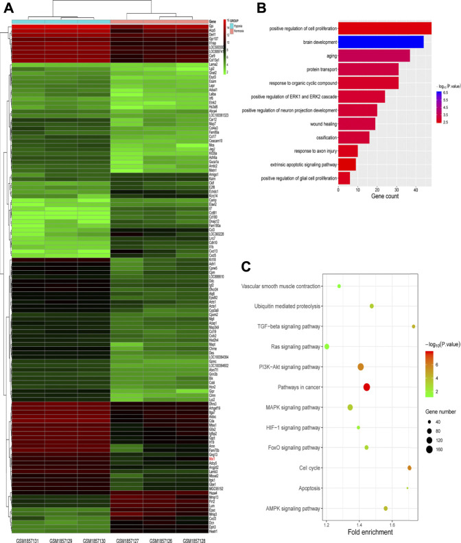
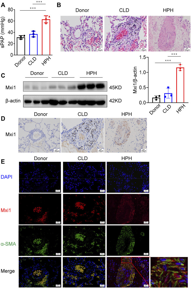
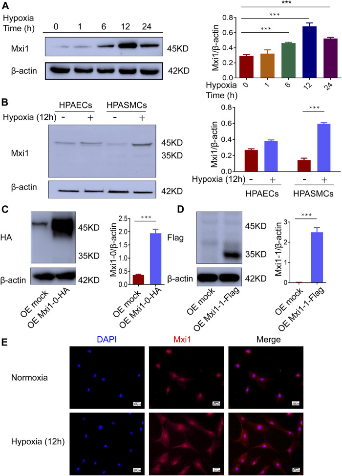
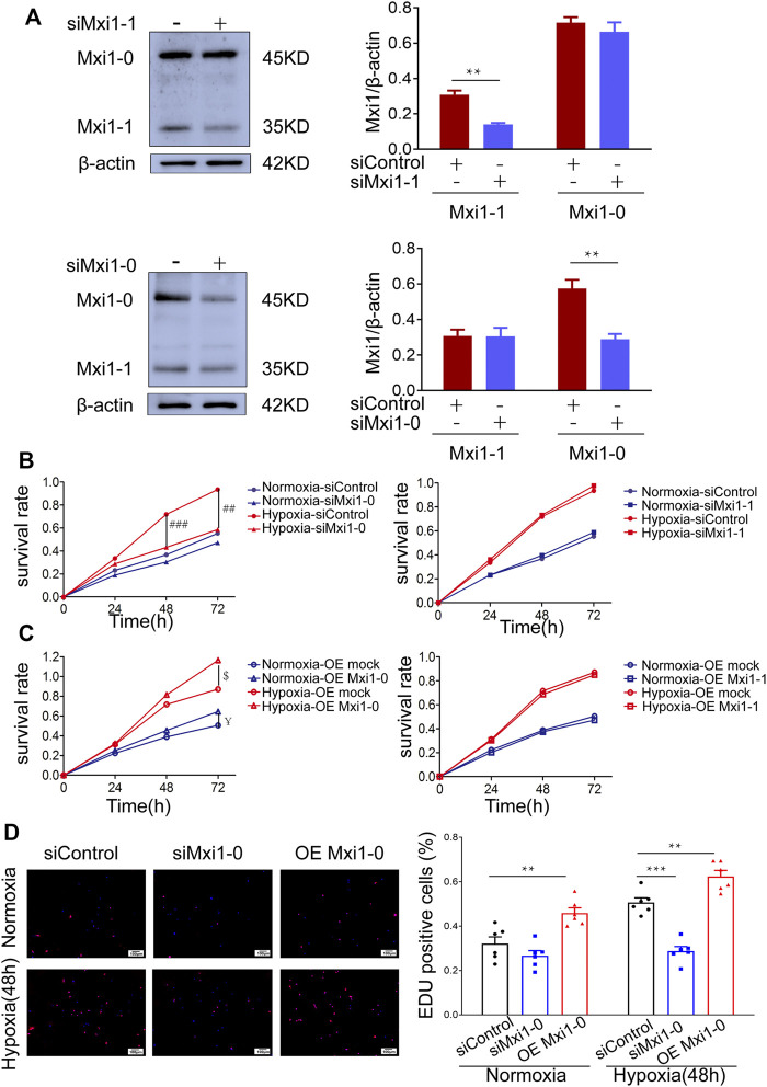
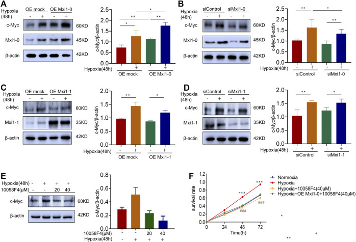
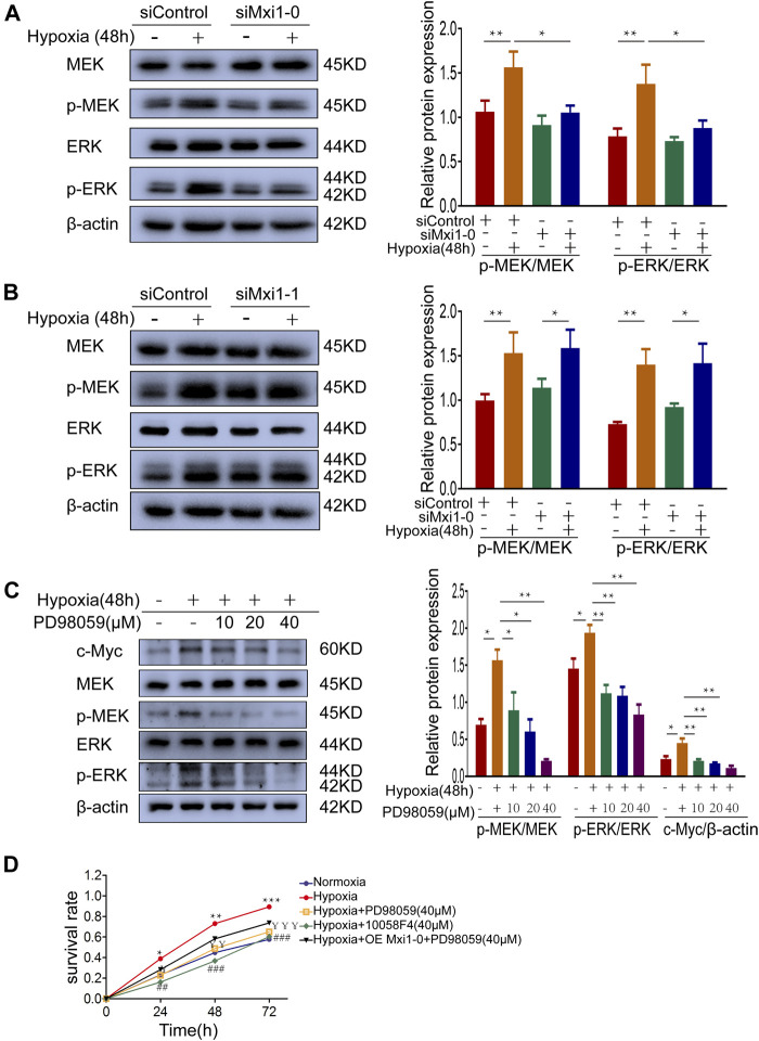
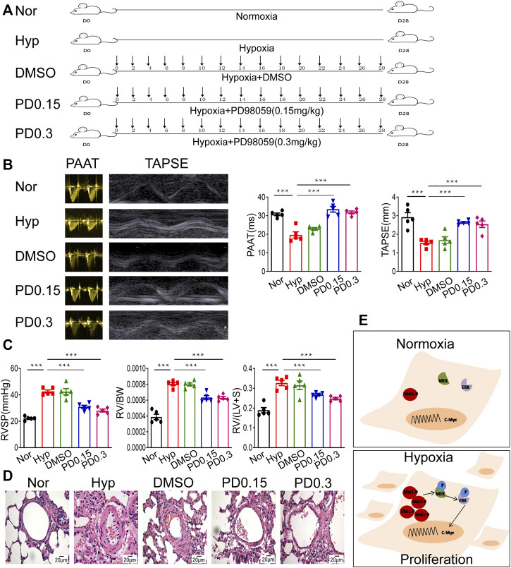
Background: Hypoxic pulmonary hypertension (HPH) is a challenging lung arterial disorder with remarkably high incidence and mortality, and so far patients have failed to benefit from therapeutics clinically available. Max interacting protein 1-0 (Mxi1-0) is one of the functional isoforms of Mxi1. Although it also binds to Max, Mxi1-0, unlike other Mxi1 isoforms, cannot antagonize the oncoprotein c-Myc because of its unique proline rich domain (PRD). While Mxi1-0 was reported to promote cell proliferation via largely uncharacterized mechanisms, it is unknown whether and how it plays a role in the pathogenesis of HPH. Methods: GEO database was used to screen for genes involved in HPH development, and the candidate players were validated through examination of gene expression in clinical HPH specimens. The effect of candidate gene knockdown or overexpression on cultured pulmonary arterial cells, e.g., pulmonary arterial smooth muscle cells (PASMCs), was then investigated. The signal pathway(s) underlying the regulatory role of the candidate gene in HPH pathogenesis was probed, and the outcome of targeting the aforementioned signaling was evaluated using an HPH rat model. Results: Mxi1 was significantly upregulated in the PASMCs of HPH patients. As the main effector isoform responding to hypoxia, Mxi1-0 functions in HPH to promote PASMCs proliferation. Mechanistically, Mxi1-0 improved the expression of the proto-oncogene c-Myc via activation of the MEK/ERK pathway. Consistently, both a MEK inhibitor, PD98059, and a c-Myc inhibitor, 10058F4, could counteract Mxi1-0-induced PASMCs proliferation. In addition, targeting the MEK/ERK signaling significantly suppressed the development of HPH in rats. Conclusion: Mxi1-0 potentiates HPH pathogenesis through MEK/ERK/c-Myc-mediated proliferation of PASMCs, suggesting its applicability in targeted treatment and prognostic assessment of clinical HPH.

Keywords: MEK/ERK; c-Myc; cell proliferation; hypoxic pulmonary hypertension (HPH); max interacting protein 1–0 (Mxi1-0); pulmonary arterial smooth muscle cells (PASMCs).


Conflict of interest statement

The authors declare that the research was conducted in the absence of any commercial or financial relationships that could be construed as a potential conflict of interest.

Figures

FIGURE 1
FIGURE 1
Screening for hypoxic pulmonary hypertension (HPH)-related genes. (A) Heatmap of genes differentially expressed in normal and hypoxic pulmonary arterial smooth muscle cells (PASMCs) in GEO database (dataset: GSM1857126∼1857131). (B,C) GO (B) and KEGG (C) enrichment analyses for differentially expressed genes described in (A).
FIGURE 2
FIGURE 2
Mxi1 is upregulated in PASMCs of HPH patients. Lung samples of three donors, three chronic lung disease (CLD) patients and three hypoxic pulmonary hypertension (HPH) patients were collected and sectioned. (A) Hemodynamic analysis of patients before collection of samples. (B) Hematoxylin and eosin staining of paraffin-fixed lung sections was used for morphological analysis of pulmonary arteries. (C) The expression of Mxi1 was examined via Western blotting analysis, followed by quantification of the blots through densitometry and normalization to β-actin. (D) Representative paraffin lung tissue sections from donors, CLD, and HPH patients were subjected to immunohistochemical staining of Mxi1. (E) Representative sections in all groups were also subjected to immunofluorescence staining of Mxi1 and α-SMA with nuclei counterstained by DAPI (blue), and the image of HPH merge was zoomed. Scale bar, 20 μm. Data are shown as means ± SDs. For statistical significance, ***represents p < 0.001 compared with HPH patients.
FIGURE 3
FIGURE 3
Hypoxia induces expression of Mxi1-0 but not Mxi1-1 in PASMCs. (A) PASMCs were exposed to hypoxia (1% O2) for indicated periods of time. Cell lysates were prepared and subjected to Western blotting assay. (B) Pulmonary arterial endothelial cells (PAECs) and PASMCs were exposed to normoxia (21% O2) or hypoxia for 12 h, and were subjected to Western blottinganalyses. (C,D) PASMCs were transfected with constructs for HA-tagged Mxi1-0 (C) or Flag-tagged Mxi1-1 (D), and were subjected to Western blotting analyses. (E) PASMCs were exposed to normoxia or hypoxia for 12 h, and were subjected to immunostaining with a Mxi1 antibody and counterstaining of the nuclei with DAPI. Scale bar, 20 μm. Data from three independent experiments are shown as means ± SDs. For statistical significance, ***represents p < 0.001 compared to normoxia or the mock-transfected group.
FIGURE 4
FIGURE 4
Mxi1-0 but not Mxi1-1 promotes PASMCs growth under hypoxic conditions. (A) PASMCs were transfected with siRNAs targeting either Mxi1-0 or Mxi1-1, and were subjected to Western blotting analyses. (B) PASMCs cultured in normoxic or hypoxic conditions were transfected with siRNAs targeting either Mxi1-0 or Mxi1-1, and CCK-8 assays were performed on indicated times. (C) PASMCs cultured in normoxic or hypoxic conditions were infected with recombinant lentiviruses to overexpress Mxi1-0 or Mxi1-1, and CCK-8 assays were performed on indicated times. (D) PASMCs were transfected with Mxi1-0-targeted siRNAs or infected with recombinant lentiviruses to overexpress Mxi1-0, and were cultured in normoxic or hypoxic conditions for 48 h. Cells were then subjected to immunofluorescence staining for EdU, followed by microscopy and calculation of the ratios of EdU-positive cells in three random fields. Scale bar, 100 μm. Data from three independent experiments are shown as means ± SDs. For statistical significance, *represents p < 0.05 compared to siControl, **represents p < 0.01 compared to siControl, ***represents p < 0.001 compared to siControl, ## represents p < 0.01 compared to Hypoxia-siControl, ### represents p < 0.001 compared to Hypoxia-siControl, $represents p < 0.05 compared to Hypoxia-OEmock, ¥represents p < 0.05 compared to Normoxia-OEmock.
FIGURE 5
FIGURE 5
Mxi1-0 upregulates c-Myc production in hypoxic PASMCs. (A) PASMCs were infected with recombinant lentiviruses to overexpress Mxi1-0, and were cultured under normoxic or hypoxic conditions for 48 h. Cells were then subjected to Western blotting analysis. (B) PASMCs were transfected with Mxi1-0-targeted siRNAs, and were cultured under normoxic or hypoxic conditions for 48 h. Cells were then subjected to Western blotting analysis. (C) PASMCs were infected with recombinant lentiviruses to overexpress Mxi1-1, and were cultured under normoxic or hypoxic conditions for 48 h. Cells were then subjected to Western blotting analysis. (D) PASMCs were transfected with Mxi1-1-targeted siRNAs, and were cultured under normoxic or hypoxic conditions for 48 h. Cells were then subjected to Western blotting analysis. (E) PASMCs were cultured under normoxic or hypoxic conditions for 48 h and treated with indicated doses of 10058F4. Cells were then subjected to Western blotting analysis. (F) PASMCs were cultured under normoxic or hypoxic conditions, treated with 10058F4, and infected with control or Mxi1-0-overexpressing lentiviruses. Cells were then subjected to CCK-8 assays on indicated times. Data from three independent experiments are shown as means ± SDs. For statistical significance, *represents p < 0.05 compared between two groups, **represents p < 0.01 between two groups, ***represents p < 0.001 compared between Hypoxia and Normoxia, ### represents p < 0.001 compared between Hypoxia and Hypoxia + 10058F4 (40 μM).
FIGURE 6
FIGURE 6
Mxi1-0 upregulates c-Myc through MEK/ERK signaling in hypoxic PASMCs. (A,B) PASMCs tranfected with siRNAs targeting Mxi1-0 (A) or Mxi1-1 (B) were cultured under normoxic or hypoxic conditions for 48 h. Western blotting assay was performed with the indicated antibodies. All the phospho-protein levels were measured by densitometry and normalized to that of β-actin. (C) PASMCs were cultured under normoxic or hypoxic conditions for 48 h and treated with different doses of PD98059, and the levels of indicated proteins were determined by Western blotting analysis and quantified by densitometry. (D) PASMCs were cultured under normoxic or hypoxic conditions, treated with PD98059 or 10058F4, and infected with control or Mxi1-0-overexpressing lentiviruses. Cells were then subjected to CCK-8 assays on indicated times. Data from three independent experiments are shown as means ± SDs. For statistical significance, *represents p < 0.05 compared between two groups or to Normoxia (D), **represents p < 0.01 compared between two groups or to Normoxia (D), ¥represents p < 0.05 compared between Hypoxia and Hypoxia + PD98059 (40 μM), ¥¥represents p < 0.01 compared between Hypoxia and Hypoxia + PD98059 (40 μM),¥¥¥represents p < 0.001 compared between Hypoxia and Hypoxia + PD98059 (40 μM), ## represents p < 0.01 compared between Hypoxia and Hypoxia+10058F4 (40 μM), ###represents p < 0.001 compared between Hypoxia and Hypoxia + 10058F4 (40 μM).
FIGURE 7
FIGURE 7
Inhibition of MEK/ERK signaling protects rats against HPH. (A) A rat model of hypoxic pulmonary hypertension (HPH) was generated (n = 5 animals per group). Rats exposed to chronic hypoxia were treated with vehicle (DMSO) or 0.15 mg/kg or 0.3 mg/kg PD98059. (B) Rats in all groups were subjected to echocardiography and measurement of PAATand TAPSE. (C) After intubation for rats described in (A), RVSP was recorded, and the right ventricular hypertrophy ratio of RV/BW and RV/LV + S were calculated. (D) Hematoxylin and eosin staining of paraffin-fixed lung sections prepared from rats described in (A) was performed for morphological analysis of the pulmonary arteries. (E) A diagram showing that hypoxia-induced Mxi1-0 promotes PASMCs proliferation via MEK/ERK/c-Myc signaling in the context of HPH. Scale bar, 20 μm. Data are shown as means ± SDs. For statistical significance, ***represents p < 0.001 compared to hypoxia. PAAT, pulmonary artery acceleration time; TAPSE, tricuspid annulusplain systolic excursion; RVSP, right ventricular systolic pressure; RV, right ventricle; BW, body weight; LV + S, left ventricle plus septum.

References

    1. Armstrong M. B., Mody R. J., Ellis D. C., Hill A. B., Erichsen D. A., Wechsler D. S. (2013). N-myc Differentially Regulates Expression of MXI1 Isoforms in Neuroblastoma. Neoplasia 15 (12), 1363–1370. PubMed PMID: 24403858; PubMed Central PMCID: PMC3884527. 10.1593/neo.11606 - DOI - PMC - PubMed
    1. Badesch D. B., Raskob G. E., Elliott C. G., Krichman A. M., Farber H. W., Frost A. E., et al. (2010). Pulmonary Arterial Hypertension. Chest 137 (2), 376–387. PubMed PMID: 19837821. 10.1378/chest.09-1140 - DOI - PubMed
    1. Baluapuri A., Wolf E., Eilers M. (2020). Target Gene-independent Functions of MYC Oncoproteins. Nat. Rev. Mol. Cel Biol 21 (5), 255–267. PubMed PMID: 32071436; PubMed Central PMCID: PMC7611238. 10.1038/s41580-020-0215-2 - DOI - PMC - PubMed
    1. Boult J. K. R., Tanière P., Hallissey M. T., Campbell M. J., Tselepis C. (2008). Oesophageal Adenocarcinoma Is Associated with a Deregulation in the MYC/MAX/MAD Network. Br. J. Cancer 98 (12), 1985–1992. PubMed PMID: 18493233; PubMed Central PMCID: PMC2441969. 10.1038/sj.bjc.6604398 - DOI - PMC - PubMed
    1. Cassady S. J., Reed R. M. (2019). Pulmonary Hypertension in COPD: A Case Study and Review of the Literature. Medicina 55 (8), 432. PubMed PMID: 31382489; PubMed Central PMCID: PMC6723523. 10.3390/medicina55080432 - DOI - PMC - PubMed