. 2022 Mar 23:13:851384.
doi: 10.3389/fgene.2022.851384. eCollection 2022.

A Novel Pyroptosis-Related Gene Signature for Predicting Prognosis in Kidney Renal Papillary Cell Carcinoma

Affiliations

A Novel Pyroptosis-Related Gene Signature for Predicting Prognosis in Kidney Renal Papillary Cell Carcinoma

Jian Hu et al. Front Genet. .

Abstract

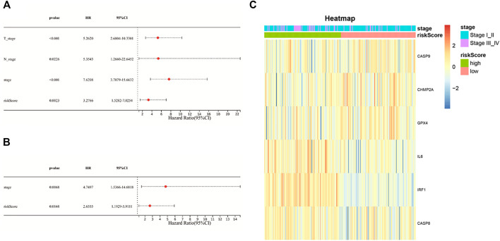
Pyroptosis is defined as an inflammatory form of programmed cell death. Increasing studies have demonstrated that pyroptosis is closely related to tumor development and antitumor process. However, the role of pyroptosis in kidney renal papillary cell carcinoma (KIRP) remains obscure. In this study, we analyzed the expression of 52 pyroptosis-related genes (PRGs) in KIRP, of which 20 differentially expressed PRGs were identified between tumor and normal tissues. Consensus clustering analysis based on these PRGs was used to divided patients into two clusters, from which a significant difference in survival was found (p = 0.0041). The prognostic risk model based on six PRGs (CASP8, CASP9, CHMP2A, GPX4, IL6, and IRF1) was built using univariate Cox regression and LASSO-Cox regression analysis, with good performance in predicting one-, three-, and five-year overall survival. Kaplan-Meier survival analysis showed that the high-risk group had a poor survival outcome (p < 0.001) and risk score was an independent prognostic factor (HR: 2.655, 95% CI 1.192-5.911, p = 0.016). Immune profiling revealed differences in immune cell infiltration between the two groups, and the infiltration of M2 macrophages was significantly upregulated in the tumor immune microenvironment, implying that tumor immunity participated in the KIRP progression. Finally, we identified two hub genes in tumor tissues (IL6 and CASP9), which were validated in vitro. In conclusion, we conducted a comprehensive analysis of PRGs in KIRP and tried to provide a pyroptosis-related signature for predicting the prognosis.

Keywords: gene; kidney renal papillary cell carcinoma; prognosis; pyroptosis; signature.


Conflict of interest statement

The authors declare that the research was conducted in the absence of any commercial or financial relationships that could be construed as a potential conflict of interest.

Figures

FIGURE 1
FIGURE 1
(A) Heat-map of differentially expressed PRGs between the tumor and normal tissues (red color: higher expression; blue color: lower expression, all p < 0.05). (B) PPI network of 20 differentially expressed PRGs obtained from the STRING database. (C) Correlation network of 20 differentially expressed PRGs (the correlation coefficients are presented by different colors: red line, positive correlation; blue line, negative correlation).
FIGURE 2
FIGURE 2
Two KIRP clusters obtained by consensus clustering analysis based on differentially expressed PRGs. (A) Area under the cumulative distribution function (CDF) curve for k = 2–9. (B) CDF delta area for k = 2–9. (C) Tracking plot for k = 2–9. (D) Consensus clustering matrix for k = 2. (E) Kaplan–Meier survival analysis of two subgroups. (F) Heat-map of PRGs and the clinical characteristics between the two clusters.
FIGURE 3
FIGURE 3
Construction of a six-gene prognostic signature in KIRP patients. (A) Univariate cox regression analysis of 11 PRGs (p < 0.2). (B,C) LASSO regression of 11 PRGs and the tuning parameter (λ) selection cross-validation curve. (D) Distribution of risk scores for KIRP patients. (E) PCA plot based on the risk score. (F) Distribution of patient survival status according to the high-risk group and the low-risk group. (G) Kaplan–Meier curves for OS in the low- and high-risk groups. (H) ROC curves to evaluate the predictive efficiency of the risk model.
FIGURE 4
FIGURE 4
(A) Univariate Cox regression analysis of the risk score and other clinical characteristics associated with overall survival; (B) multivariate Cox regression analysis of the risk score and other clinical characteristics; (C) heat-map showing the relationship of the risk groups and tumor stage.
FIGURE 5
FIGURE 5
(A–C) GO functional enrichment analysis of DEGs in the two risk groups (BP, biological process; CC, cellular component; MF, molecular function); (D) KEGG pathway analysis of DEGs in the two risk groups.
FIGURE 6
FIGURE 6
(A,C) Differences in immune cell composition between the high-risk group and the low-risk group; (B) tumor microenvironment immune cell composition in KIRP patients.
FIGURE 7
FIGURE 7
(A) The PPI network was constructed containing six genes of the signature. (B) Screening hub genes from the PPI network (red node: genes with a high MCC score; blue node: genes with a low MCC score). (C–E) The cohort was divided into two groups (high and low) according to their median expression value separately, and the expressions of IL6, RAF1, and CASP9 were associated with overall survival (p < 0.05). (F,G) Results of IL6 and CASP9 in immuno-histochemical staining between KIRP and normal tissues (scale bar values: 100 µm).

References

    1. Block K. M., Hanke N. T., Maine E. A., Baker A. F. (2012). IL-6 Stimulates STAT3 and Pim-1 Kinase in Pancreatic Cancer Cell Lines. Pancreas 41 (5), 773–781. 10.1097/MPA.0b013e31823cdd10 - DOI - PMC - PubMed
    1. Broz P., Pelegrín P., Shao F. (2020). The Gasdermins, a Protein Family Executing Cell Death and Inflammation. Nat. Rev. Immunol. 20 (3), 143–157. 10.1038/s41577-019-0228-2 - DOI - PubMed
    1. Brozovich A., Garmezy B., Pan T., Wang L., Farach-Carson M. C., Satcher R. L. (2021). All Bone Metastases Are Not Created Equal: Revisiting Treatment Resistance in Renal Cell Carcinoma. J. Bone Oncol. 31, 100399. 10.1016/j.jbo.2021.100399 - DOI - PMC - PubMed
    1. Chavez-Dominguez R., Perez-Medina M., Aguilar-Cazares D., Galicia-Velasco M., Meneses-Flores M., Islas-Vazquez L., et al. (2021). Old and New Players of Inflammation and Their Relationship with Cancer Development. Front. Oncol. 11, 722999. 10.3389/fonc.2021.722999 - DOI - PMC - PubMed
    1. Derangère V., Chevriaux A., Courtaut F., Bruchard M., Berger H., Chalmin F., et al. (2014). Liver X Receptor β Activation Induces Pyroptosis of Human and Murine colon Cancer Cells. Cell Death Differ 21 (12), 1914–1924. 10.1038/cdd.2014.117 - DOI - PMC - PubMed