Review
. 2022 Mar 14;12(6):2833-2859.
doi: 10.7150/thno.70719. eCollection 2022.

Emulating interactions between microorganisms and tumor microenvironment to develop cancer theranostics

Affiliations
Review

Emulating interactions between microorganisms and tumor microenvironment to develop cancer theranostics

Tongmeng Jiang et al. Theranostics. .

Abstract

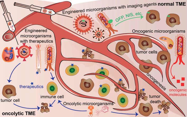
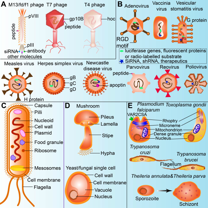
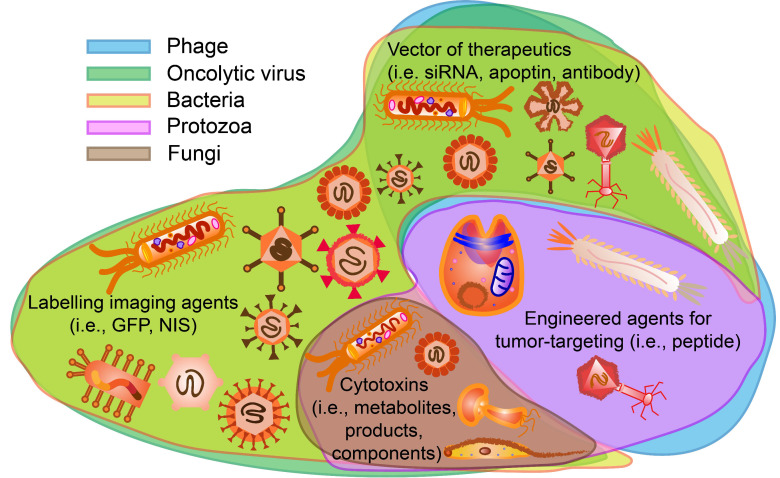
The occurrence of microorganisms has been confirmed in the tumor microenvironment (TME) of many different organs. Microorganisms (e.g., phage, virus, bacteria, fungi, and protozoa) present in TME modulate TME to inhibit or promote tumor growth in species-dependent manners due to the special physiological and pathological features of each microorganism. Such microorganism-TME interactions have recently been emulated to turn microorganisms into powerful cancer theranostic agents. To facilitate scientists to explore microorganisms-TME interactions further to develop improved cancer theranostics, here we critically review the characteristics of different microorganisms that can be found in TME, their interactions with TME, and their current applications in cancer diagnosis and therapy. Clinical trials of using microorganisms for cancer theranostics are also summarized and discussed. Moreover, the emerging technology of whole-metagenome sequencing that can be employed to precisely determine microbiota spectra is described. Such technology enables scientists to gain an in-depth understanding of the species and distributions of microorganisms in TME. Therefore, scientists now have new tools to identify microorganisms (either naturally present in or introduced into TME) that can be used as effective probes, monitors, vaccines, or drugs for potentially advancing cancer theranostics to clinical applications.

Keywords: cancer theranostics; microbiota spectra; microorganisms; tumor microenvironment (TME).


Conflict of interest statement

Competing Interests: The authors have declared that no competing interest exists.

Figures

Figure 1
Figure 1
Microorganisms existing in normal tumor microenvironment (TME) and oncolytic TME present different functions. Oncogenic microorganism expressed molecules that stimulate oncogenesis. Engineered microorganisms are designed as monitors or diagnostic factors in normal TME, and serve as monitors or therapeutic factors in oncolytic TME. Some microorganisms are acting as anti-tumoral therapeutics themselves in TME.
Figure 2
Figure 2
The unique structures of microorganisms applied for cancer theranostics. (A) Phage can be modified for displaying tumor-targeting peptides, anti-tumor agents, as well as acting as oncolytic factors themselves. (B) Oncolytic viruses can be labelled with imaging agents or therapeutics for cancer theranostics. (C) Bacteria uptake nanoparticles or imaging agents as food granule for tumor imaging. Bacterial mesosomes and ribosome contribute to excrete anti-cancerous enzymes or agents. The flagella and LPS in the bacteria cell wall can modulate immune response for cancer therapy. (D) Polysaccharides in the cell walls of fungi (including mushroom and yeast) could stimulate immune cells for cancer therapy. (E) Protozoa expressing or modified with therapeutics (ie.VAR2CSA) and flagella in the protozoa are sometimes anti-tumorous. Transformation of sporozoites into schizonts stimulate apoptosis and proinflammation.
Figure 3
Figure 3
Oligopeptides screened by phage display can be used for ovarian cancer diagnosis. Adapted with permission from , Copyright 2015, Ivyspring International Publisher, CC BY-NC 4.0.
Figure 4
Figure 4
Near-infrared fluorescence imaging probes based on M13KO7 phage display. Adapted with permission from , Copyright 2018, Ivyspring International Publisher, CC BY-NC 4.0.
Figure 5
Figure 5
Peptides screened by phage display and used for targeted cancer therapy. Adapted with permission from , Copyright 2019, Springer Nature Switzerland AG. Part of Springer Nature, CC BY 4.0.
Figure 6
Figure 6
Selection of oncolytic vaccinia virus for personalized therapy. Adapted with permission from . Copyright 2012, Ivyspring International Publisher, CC BY-NC 4.0.
Figure 7
Figure 7
Oncolytic NIS-expressing adenovirus enhances cancer imaging in pancreatic cancer models. Adapted with permission from , Copyright 2021, Elsevier, CC BY-NC-ND 4.0.
Figure 8
Figure 8
Administration of engineered adenoviruses suppresses tumor growth and prolongs survival of lung cancer bearing mice. (A) Bioluminescence images of subcutaneous tumor-bearing mice after administration of viruses. (B) Activity of viruses. (C) Amounts of viruses. (D) Tumor volume after administration of viruses. (E) Survival of subcutaneous tumor-bearing mice after administration of viruses. (F) Viral genome copies in the lungs after administration of viruses. (G) Immunofluorescent staining of lung tumors after administration of viruses. (H) Survival of orthotopic tumor-bearing mice after administration of viruses. (I) Bioluminescence images of orthotopic tumor-bearing mice after administration of viruses. (J) HE staining of lung tumor. Adapted with permission from , Copyright 2020, The American Association for the Advancement of Science.
Figure 9
Figure 9
Bacteria uptake 18F-FDS for tumor imaging by PET. Adapted with permission from , Copyright 2020, Ivyspring International Publisher, CC BY 4.0.
Figure 10
Figure 10
Anticancer effects of Listeria monocytogenes through an immune response. Adapted with permission from , Copyright 2018, MDPI, Basel, Switzerland, CC BY 4.0.
Figure 11
Figure 11
Polysaccharides from Ganoderma sinense suppress lung cancer in mice model. (A) Tumor volume. (B) H&E staining. Adapted with permission from , Copyright 2021, BioMed Central Ltd. Part of Springer Nature, CC BY 4.0.
Figure 12
Figure 12
The plasmodium expressing VAR2CSA and recombinant VAR2CSA (rVAR2) can be applied to cancer diagnosis and therapy. Adapted with permission from , Copyright 2018, Elsevier Ltd.
Figure 13
Figure 13
Trypanosoma cruzi extracts elicit protective immune response against chemically induced colon and mammary cancers. Adapted with permission from , Copyright 2015, UICC, John Wiley and Sons.
Figure 14
Figure 14
Potential functions of microorganisms on tumor cells for cancer theranostics. Phages and protozoa can display tumor-targeting agents such as peptides. Oncolytic viruses and bacteria can be labelled with imaging agents such as GFP, NIS and so on. Phages oncolytic viruses and bacteria are possible vectors for delivering certain therapeutics including apoptin, siRNA and antibodies. Oncolytic viruses, bacteria, fungi and protozoa contain or express cytotoxic components that can assist cancer therapy.
Figure 15
Figure 15
Abundance of F. nucleatum and C. symbiosum relative to colorectal cancer. Adapted with permission from . Copyright 2017, Elsevier B.V, CC BY-NC-ND 4.0.
Figure 16
Figure 16
Decreased tumor metabolic activity shown in a patient after HSV1716 administration. Adapted with permission from . Copyright 2019, Elsevier Ltd, CC BY-NC-ND 4.0.

Similar articles

Cited by

References

    1. Bray F, Ferlay J, Soerjomataram I, Siegel RL, Torre LA, Jemal A. Global cancer statistics 2018: GLOBOCAN estimates of incidence and mortality worldwide for 36 cancers in 185 countries. CA Cancer J Clin. 2018;68:394–424. - PubMed
    1. Najafi M, Goradel NH, Farhood B, Salehi E, Solhjoo S, Toolee H. et al. Tumor microenvironment: Interactions and therapy. J Cell Physiol. 2019;234:5700–21. - PubMed
    1. Jobin C. Precision medicine using microbiota. Science. 2018;359:32–4. - PubMed
    1. El-Jurdi N, Ghannoum MA. The mycobiome: Impact on health and disease states. Microbiol Spectr. 2017;5:FUNK–0045. -2016. - PMC - PubMed
    1. Dzutsev A, Goldszmid RS, Viaud S, Zitvogel L, Trinchieri G. The role of the microbiota in inflammation, carcinogenesis, and cancer therapy. Eur J Immunol. 2015;45:17–31. - PubMed

Publication types