

Direct pulp capping of carious immature tooth using bioactive material: A case report
- PMID: 35401982
- PMCID: PMC8984855
- DOI: 10.1177/2050313X221087565
Direct pulp capping of carious immature tooth using bioactive material: A case report
Abstract
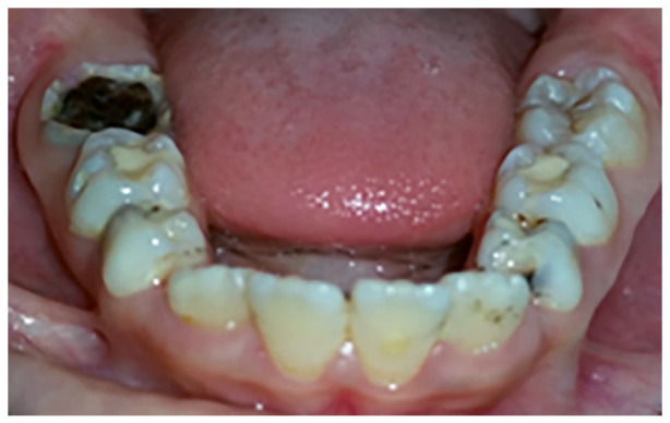
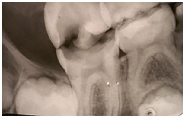
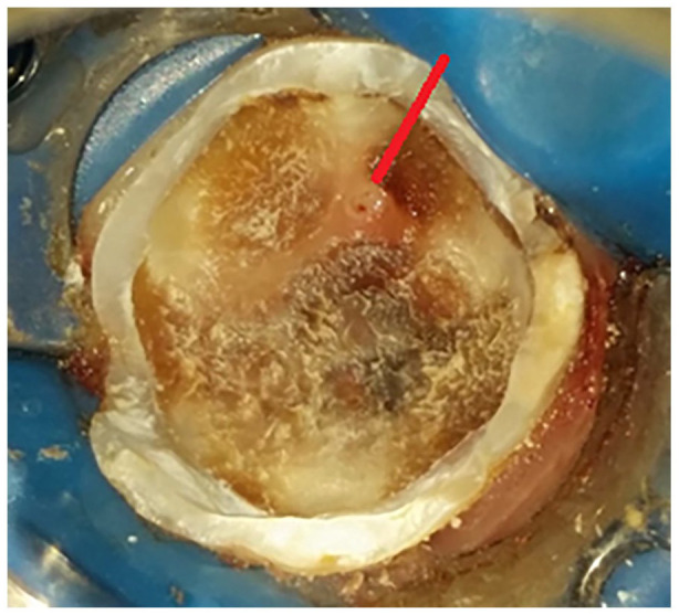
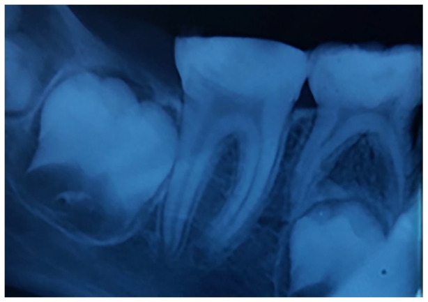
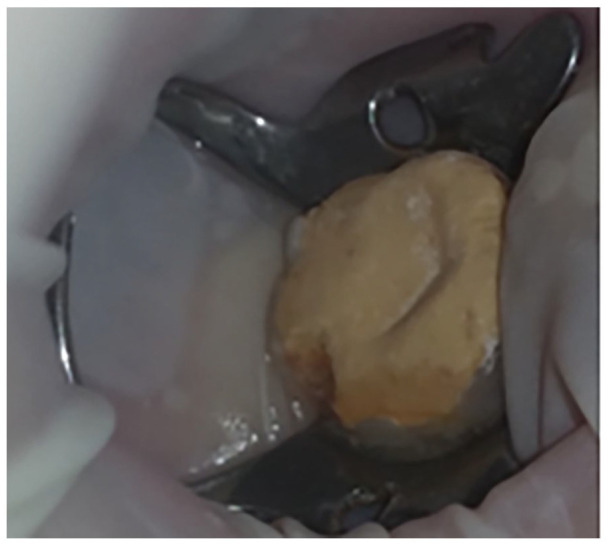
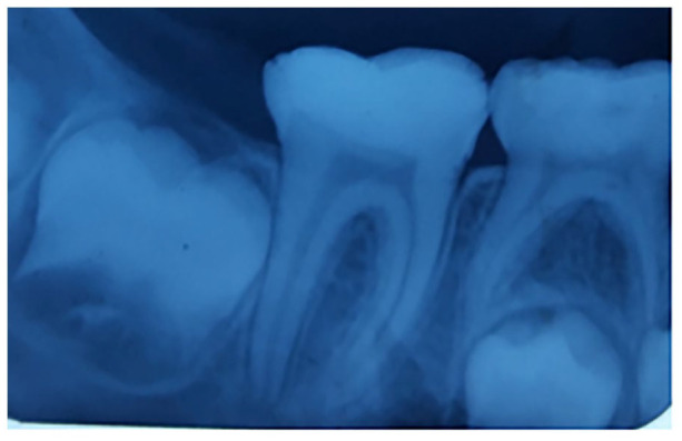
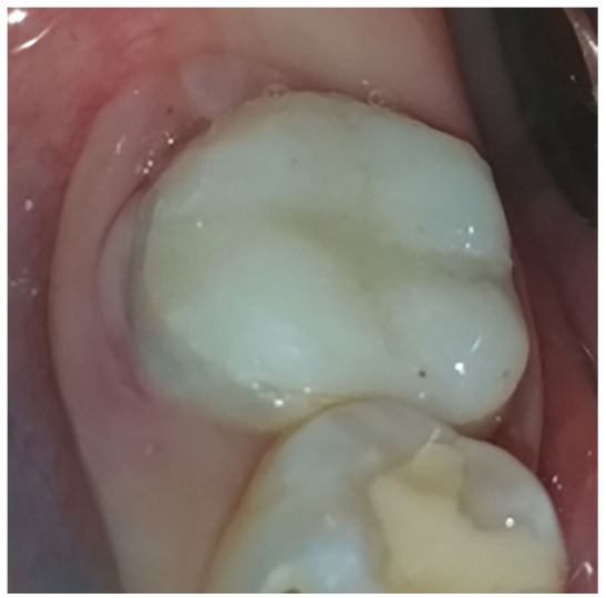
The management of deep carious lesions in immature tooth and the preservation of its pulp vitality is a real challenge in daily dental practice. Bioactive cements are of interest to deal with such cases. Our case report describes the immediate management and the follow-up of an extensive carious lesion on an immature first right mandibular molar with pulp exposure by direct pulp capping using Biodentine. A 6-month clinical and radiographic follow up showed that the tooth was vital, with dentine-bridge formation in the pulp chamber with continuous root formation. This procedure allowed the protection of pulp complex, preserving at the same time its functional and biologic activities due to the capacities of Biodentine as an effective pulp capping material to induce pulp cells to form hard tissue. The aim of this article is to discuss through the report of this clinical case, the indications, advantages and disadvantages of different procedures and biomaterials used for direct pulp capping.
Keywords: Biodentine; dentistry; direct pulp capping; permanent immature tooth; tricalcium silicate cement.
© The Author(s) 2022.
Conflict of interest statement
Declaration of conflicting interests: The author(s) declared no potential conflicts of interest with respect to the research, authorship, and/or publication of this article.
Figures
References
-
- Paula AB, Laranjo M, Marto CM, et al. Direct pulp capping: what is the most effective therapy? Systematic review and meta-analysis. J Evid Based Dent Pract 2018; 18(4): 298–314. - PubMed
Publication types
LinkOut - more resources
Full Text Sources
