

PD-L1 blockade potentiates the antitumor effects of ALA-PDT and optimizes the tumor microenvironment in cutaneous squamous cell carcinoma
- PMID: 35402079
- PMCID: PMC8986186
- DOI: 10.1080/2162402X.2022.2061396
PD-L1 blockade potentiates the antitumor effects of ALA-PDT and optimizes the tumor microenvironment in cutaneous squamous cell carcinoma
Abstract
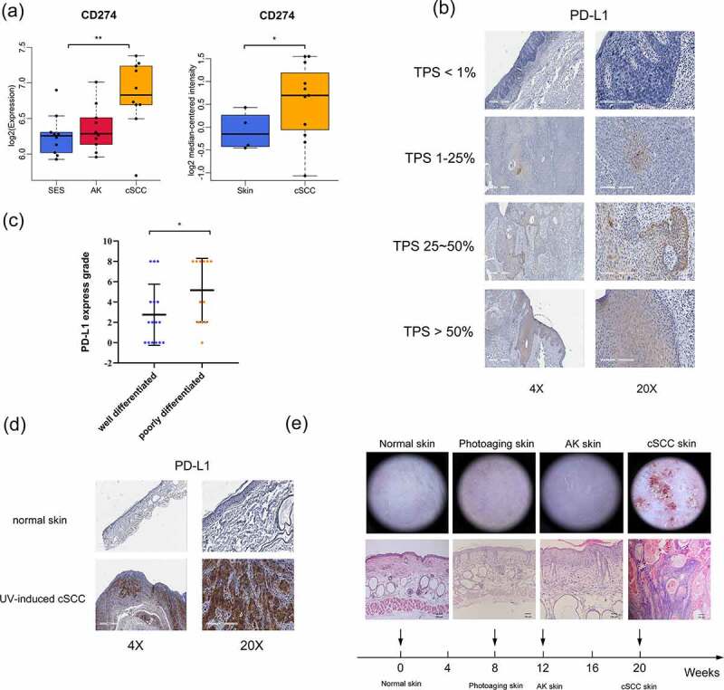
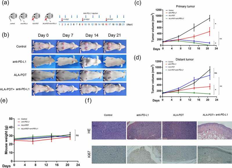
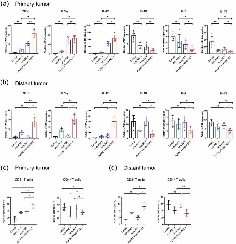
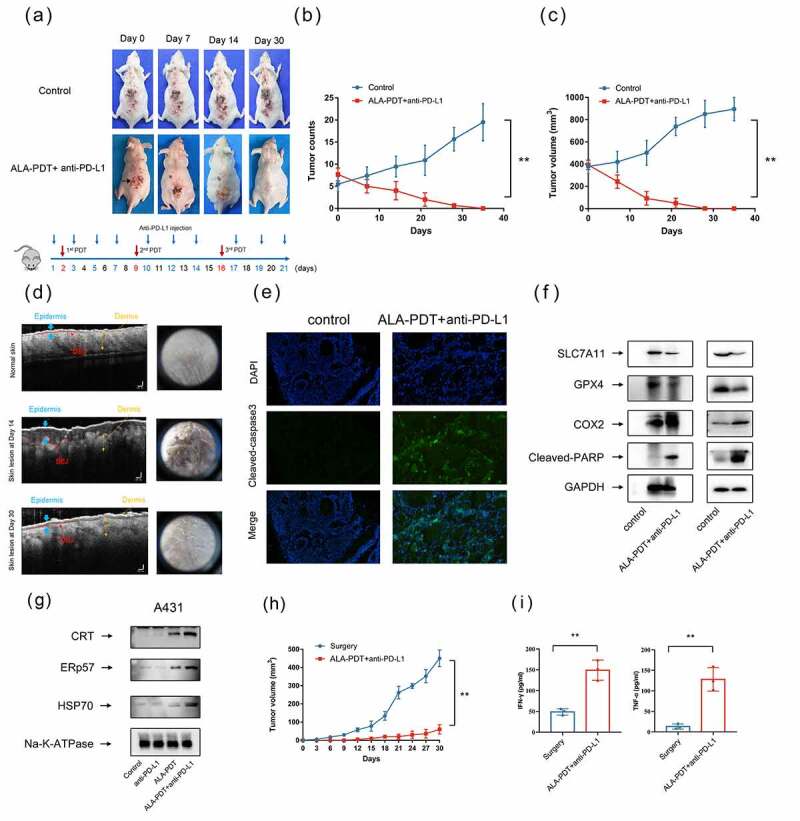
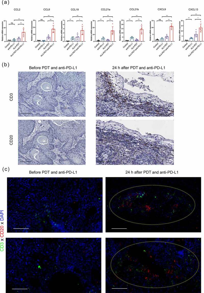
Immune checkpoint blockade (ICB) is a powerful oncologic treatment modality for a wide variety of human malignancies, but the patient response rate to this treatment remains low, especially in patients with cutaneous squamous cell carcinoma (cSCC). 5-Aminoleuvulinic acid-photodynamic therapy (ALA-PDT) is widely used to treat cancerous and precancerous skin diseases, but the value of ALA-PDT in the treatment of invasive cSCC is debatable. Our previous studies have shown that ALA-PDT can induce antitumor immune responses by promoting the immunogenic death of tumor cells. However, it is unclear whether ALA-PDT exerts synergistic effects with ICB in cSCC. Here, we report that PD-L1 blockade potentiates the antitumor effects of ALA-PDT both on primary and distant tumors, and optimizes the tumor microenvironment in cSCC. In this study, we first detected PD-L1 expression in patients with different grades of cSCC. Then we found the combination of anti-PD-L1 monoclonal antibody (mAb) and ALA-PDT killed tumor cells by apoptosis- and/or ferroptosis-mediated immunogenic cell death (ICD) and stimulated systemic immune response, as well as building the immunological memory response to prevent tumor recurrence. Furthermore, we found that combination therapy can be used to recruit tertiary lymphoid structure (TLS)-like intratumoral lymphoid aggregates, which may promote tumor-infiltrating lymphocyte (TIL)-mediated antitumor immunity. In summary, our work demonstrates that ICB treatment with an anti-PD-L1 antibody is a promising strategy that may potentiate the antitumor effects of ALA-PDT in cSCC.
Keywords: 5-aminolevulinic acid photodynamic therapy; PD-L1; cutaneous squamous cell carcinoma.
© 2022 The Author(s). Published with license by Taylor & Francis Group, LLC.
Conflict of interest statement
The authors declare no conflict of interest in this work.
Figures

Similar articles
-
CCL8 enhances sensitivity of cutaneous squamous cell carcinoma to photodynamic therapy by recruiting M1 macrophages.Photodiagnosis Photodyn Ther. 2019 Jun;26:235-243. doi: 10.1016/j.pdpdt.2019.03.014. Epub 2019 Mar 19. Photodiagnosis Photodyn Ther. 2019. PMID: 30902794
-
The effect of C-X-C motif chemokine ligand 13 in cutaneous squamous cell carcinoma treated with aminolevulinic acid-photodynamic therapy.Photodiagnosis Photodyn Ther. 2019 Jun;26:389-394. doi: 10.1016/j.pdpdt.2019.04.018. Epub 2019 Apr 22. Photodiagnosis Photodyn Ther. 2019. PMID: 31022580
-
In situ immunogenic clearance induced by a combination of photodynamic therapy and rho-kinase inhibition sensitizes immune checkpoint blockade response to elicit systemic antitumor immunity against intraocular melanoma and its metastasis.J Immunother Cancer. 2021 Jan;9(1):e001481. doi: 10.1136/jitc-2020-001481. J Immunother Cancer. 2021. PMID: 33479026 Free PMC article.
-
Immune Checkpoint Blockade in Advanced Cutaneous Squamous Cell Carcinoma: What Do We Currently Know in 2020?Int J Mol Sci. 2020 Dec 6;21(23):9300. doi: 10.3390/ijms21239300. Int J Mol Sci. 2020. PMID: 33291277 Free PMC article. Review.
-
The Next Immune-Checkpoint Inhibitors: PD-1/PD-L1 Blockade in Melanoma.Clin Ther. 2015 Apr 1;37(4):764-82. doi: 10.1016/j.clinthera.2015.02.018. Epub 2015 Mar 29. Clin Ther. 2015. PMID: 25823918 Free PMC article. Review.
Cited by
-
A review on ferroptosis and photodynamic therapy synergism: Enhancing anticancer treatment.Heliyon. 2024 Mar 31;10(7):e28942. doi: 10.1016/j.heliyon.2024.e28942. eCollection 2024 Apr 15. Heliyon. 2024. PMID: 38601678 Free PMC article. Review.
-
Ce6 derivative photodynamic therapy triggers PANoptosis and enhances antitumor immunity with LAG3 blockade in cutaneous squamous cell carcinoma.Cell Rep Med. 2025 Jul 15;6(7):102239. doi: 10.1016/j.xcrm.2025.102239. Cell Rep Med. 2025. PMID: 40669448 Free PMC article.
-
Prodrugs of paclitaxel improve in situ photo-vaccination.Photochem Photobiol. 2024 Oct 9:10.1111/php.14025. doi: 10.1111/php.14025. Online ahead of print. Photochem Photobiol. 2024. PMID: 39384406
-
Photodynamic Therapy Combined with Ferroptosis Is a Synergistic Antitumor Therapy Strategy.Cancers (Basel). 2023 Oct 19;15(20):5043. doi: 10.3390/cancers15205043. Cancers (Basel). 2023. PMID: 37894410 Free PMC article. Review.
-
Trial watch: chemotherapy-induced immunogenic cell death in oncology.Oncoimmunology. 2023 Jun 3;12(1):2219591. doi: 10.1080/2162402X.2023.2219591. eCollection 2023. Oncoimmunology. 2023. PMID: 37284695 Free PMC article. Review.
References
-
- Grob JJ, Gonzalez R, Basset-Seguin N, Vornicova O, Schachter J, Joshi A, Meyer N, Grange F, Piulats JM, Bauman JR, et al. Pembrolizumab Monotherapy for Recurrent or Metastatic Cutaneous Squamous Cell Carcinoma: a Single-Arm Phase II Trial (KEYNOTE-629. J Clin Oncol. 2020;38(25):2916–2925. doi:10.1200/JCO.19.03054. - DOI - PMC - PubMed
Publication types
MeSH terms
Substances
LinkOut - more resources
Full Text Sources
Medical
Research Materials