

Hydrocele of the Canal of Nuck: A Review
- PMID: 35402114
- PMCID: PMC8980195
- DOI: 10.7759/cureus.23757
Hydrocele of the Canal of Nuck: A Review
Abstract
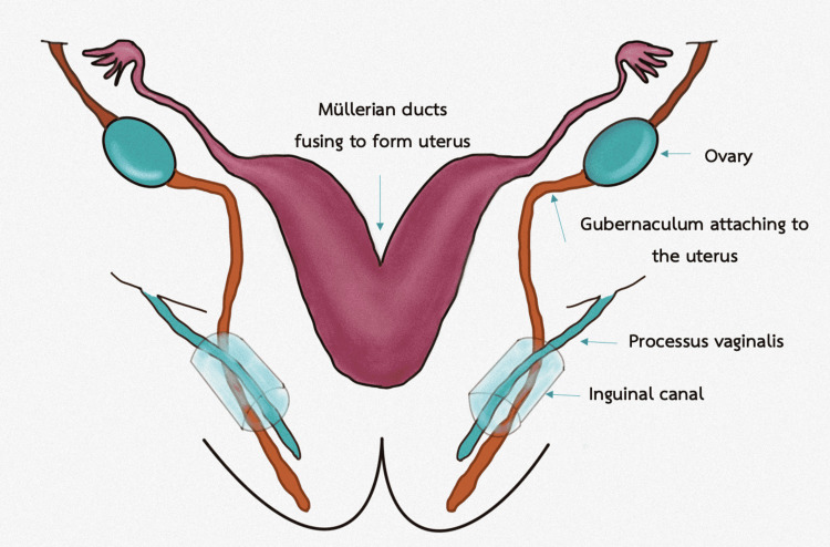
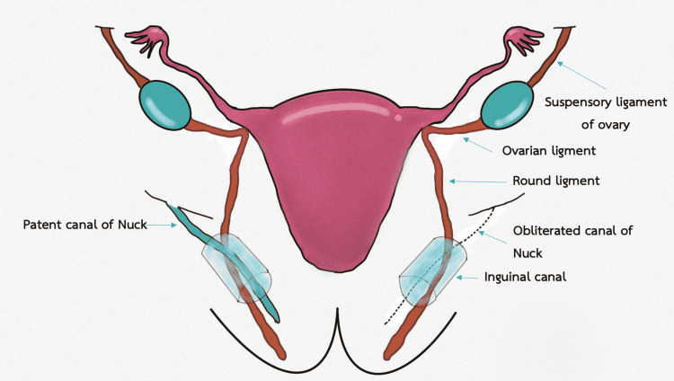
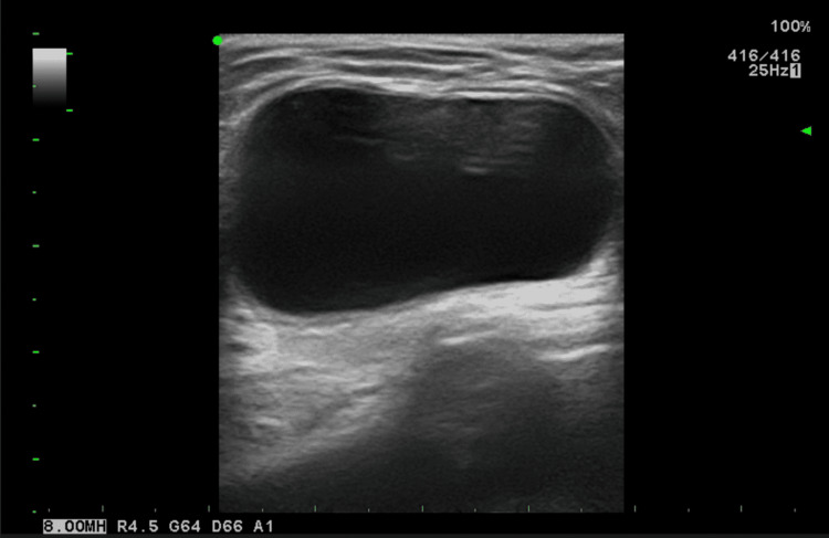
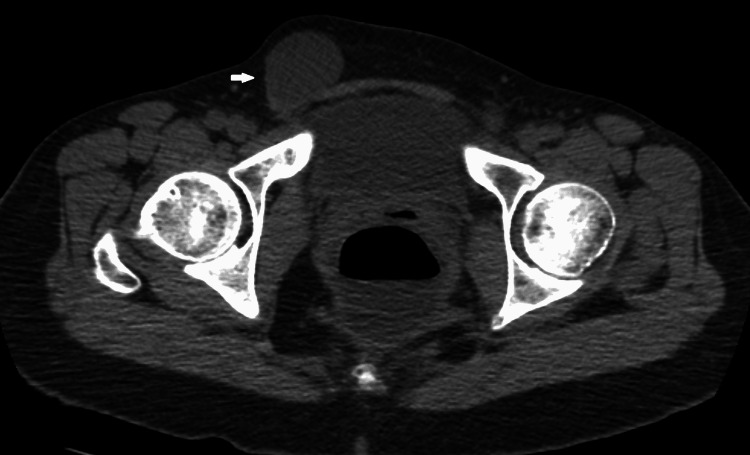
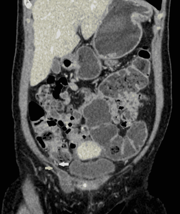
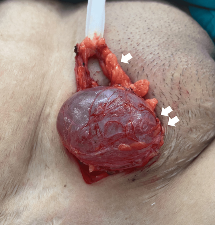
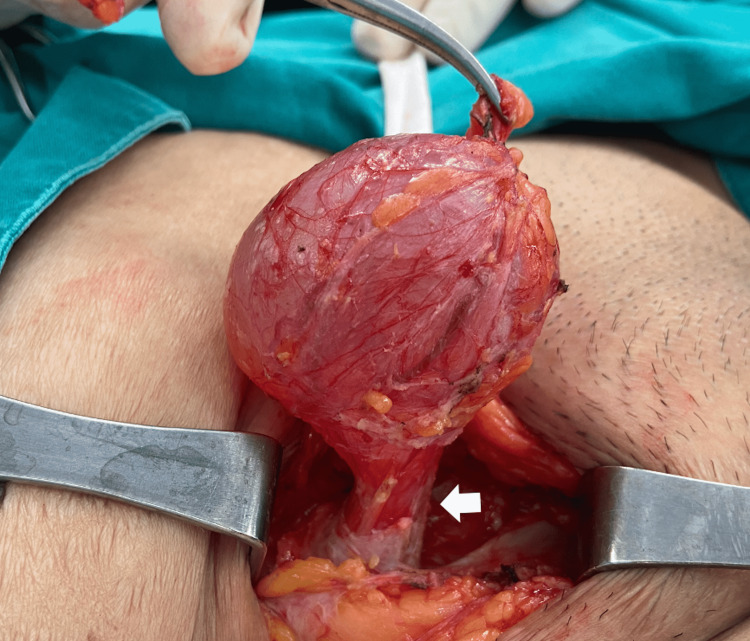
Canal of Nuck abnormality is a rare surgical condition. The pathologies are mostly encountered in young girls, less than five years of age. The incidence is even less in adults. Various pathologic conditions related to the failure of processus vaginalis obliteration can occur, involving herniation of intraabdominal structures including intestinal and genital contents such as the uterus, fallopian tube, and ovary and hydrocele of the canal of Nuck. According to its rarity, hydrocele of canal of Nuck is often misdiagnosed for common groin masses. This review summarizes and simplifies embryology, the pathophysiology of the canal of Nuck abnormalities, imaging findings, and treatment options with emphasis on the hydrocele.
Keywords: hernia; hydrocele; hydrocele of the canal of nuck; inguinal mass; the canal of nuck.
Copyright © 2022, Keeratibharat et al.
Conflict of interest statement
The authors have declared that no competing interests exist.
Figures
References
-
- Anatomy and pathology of the canal of Nuck. Nasser H, King M, Rosenberg HK, Rosen A, Wilck E, Simpson WL. Clin Imaging. 2018;51:83–92. - PubMed
-
- Imaging of groin masses: inguinal anatomy and pathologic conditions revisited. Shadbolt CL, Heinze SB, Dietrich RB. Radiographics. 2001;21 Spec No:0. - PubMed
-
- How embryology knowledge can help radiologists in the differential diagnosis of canal of Nuck pathologies. Rosa F, Martinetti C, Veirana MA, et al. Radiol Med. 2021;126:910–924. - PubMed
Publication types
LinkOut - more resources
Full Text Sources