

Expansion of Intestinal Secretory Cell Population Induced by Listeria monocytogenes Infection: Accompanied With the Inhibition of NOTCH Pathway
- PMID: 35402308
- PMCID: PMC8990097
- DOI: 10.3389/fcimb.2022.793335
Expansion of Intestinal Secretory Cell Population Induced by Listeria monocytogenes Infection: Accompanied With the Inhibition of NOTCH Pathway
Abstract
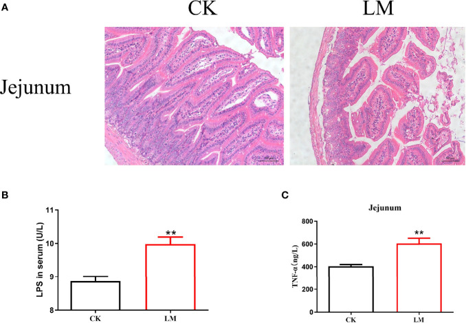
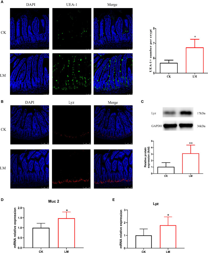
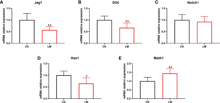
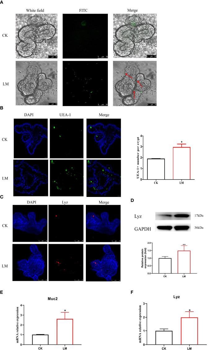
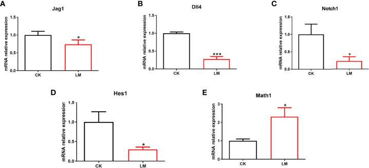
Listeria monocytogenes, as a model organism, is a causative agent of enteric pathogen that causes systemic infection. However, the interaction of L. monocytogenes and small intestinal epithelium has not been fully elucidated yet. In this study, mice and intestinal organoids were chosen as the models to investigate the influence of L. monocytogenes infection on the intestinal secretory cells and its differentiation-related pathways. Results confirmed the phenomenon of intestinal damage that L. monocytogenes infection could lead to villi damage in mice, which was accompanied by the increase of TNF-α production in jejunum as well as lipopolysaccharide (LPS) secretion in serum. Moreover, it was demonstrated that L. monocytogenes infection increased the number of goblet and Paneth cells in mice and intestinal organoids and upregulated the expression of Muc2 and Lyz. Furthermore, L. monocytogenes decreased the relative expression of Notch pathway-related genes (Jag1, Dll4, Notch1, and Hes1) while upregulating the relative expression of Math1 gene in mice and intestinal organoids. This indicated that L. monocytogenes infection caused the inhibition of Notch pathway, which may be the reason for the increased number of goblet and Paneth cells in the intestine. Collectively, these results are expected to provide more information on the mechanism of L. monocytogenes infection in the intestine.
Keywords: Listeria monocytogenes; Paneth cell; goblet cell; intestine; notch pathway.
Copyright © 2022 Zhou, Zhang, Bassey, Huang, Zou and Ye.
Conflict of interest statement
The authors declare that the research was conducted in the absence of any commercial or financial relationships that could be construed as a potential conflict of interest.
Figures
Similar articles
-
Effect of Listeria monocytogenes on intestinal stem cells in the co-culture model of small intestinal organoids.Microb Pathog. 2021 Apr;153:104776. doi: 10.1016/j.micpath.2021.104776. Epub 2021 Feb 3. Microb Pathog. 2021. PMID: 33548482
-
Increased Listeria monocytogenes Dissemination and Altered Population Dynamics in Muc2-Deficient Mice.Infect Immun. 2021 Mar 17;89(4):e00667-20. doi: 10.1128/IAI.00667-20. Print 2021 Mar 17. Infect Immun. 2021. PMID: 33431704 Free PMC article.
-
Intestinal and splenic T cell responses to enteric Listeria monocytogenes infection: distinct repertoires of responding CD8 T lymphocytes.J Immunol. 2001 Mar 15;166(6):4065-73. doi: 10.4049/jimmunol.166.6.4065. J Immunol. 2001. PMID: 11238655
-
Listeria as an enteroinvasive gastrointestinal pathogen.Curr Top Microbiol Immunol. 2009;337:173-95. doi: 10.1007/978-3-642-01846-6_6. Curr Top Microbiol Immunol. 2009. PMID: 19812983 Review.
-
Tracing innate immune defences along the path of Listeria monocytogenes infection.Immunol Cell Biol. 2014 Aug;92(7):563-9. doi: 10.1038/icb.2014.27. Epub 2014 Apr 15. Immunol Cell Biol. 2014. PMID: 24732075 Review.
Cited by
-
Current methodologies available to evaluate the virulence potential among Listeria monocytogenes clonal complexes.Front Microbiol. 2024 Oct 10;15:1425437. doi: 10.3389/fmicb.2024.1425437. eCollection 2024. Front Microbiol. 2024. PMID: 39493856 Free PMC article. Review.
-
Applications of human organoids in the personalized treatment for digestive diseases.Signal Transduct Target Ther. 2022 Sep 27;7(1):336. doi: 10.1038/s41392-022-01194-6. Signal Transduct Target Ther. 2022. PMID: 36167824 Free PMC article. Review.
-
Organoids in gastrointestinal diseases: from bench to clinic.MedComm (2020). 2024 Jun 29;5(7):e574. doi: 10.1002/mco2.574. eCollection 2024 Jul. MedComm (2020). 2024. PMID: 38948115 Free PMC article. Review.
-
Direct and indirect effects of pathogenic bacteria on the integrity of intestinal barrier.Therap Adv Gastroenterol. 2023 May 30;16:17562848231176427. doi: 10.1177/17562848231176427. eCollection 2023. Therap Adv Gastroenterol. 2023. PMID: 37274298 Free PMC article. Review.
-
The role of the Notch signalling pathway in the pathogenesis of ulcerative colitis: from the perspective of intestinal mucosal barrier.Front Med (Lausanne). 2024 Jan 5;10:1333531. doi: 10.3389/fmed.2023.1333531. eCollection 2023. Front Med (Lausanne). 2024. PMID: 38249980 Free PMC article. Review.
References
-
- Alkhuriji A. F., Majrashi N. A., Alomar S., El-Khadragy M. F., Awad M. A., Khatab A. R., et al. . (2020). The Beneficial Effect of Eco-Friendly Green Nanoparticles Using Garcinia Mangostana Peel Extract Against Pathogenicity of Listeria Monocytogenes in Female BALB/C Mice. Animals 10 (4), 573. doi: 10.3390/ani10040573 - DOI - PMC - PubMed
Publication types
MeSH terms
LinkOut - more resources
Full Text Sources
Medical
Miscellaneous