Review
. 2022 Mar 25:10:856398.
doi: 10.3389/fbioe.2022.856398. eCollection 2022.

Biodegradable Inks in Indirect Three-Dimensional Bioprinting for Tissue Vascularization

Affiliations
Review

Biodegradable Inks in Indirect Three-Dimensional Bioprinting for Tissue Vascularization

Yiting Ze et al. Front Bioeng Biotechnol. .

Abstract

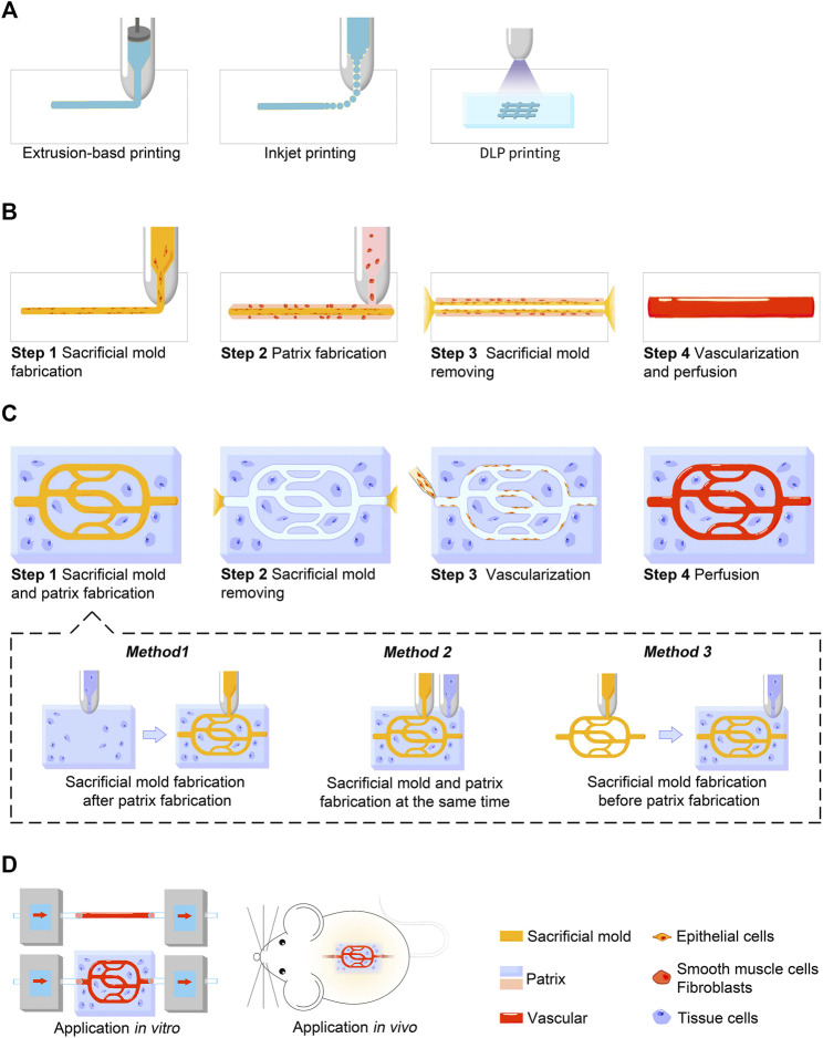
Mature vasculature is important for the survival of bioengineered tissue constructs, both in vivo and in vitro; however, the fabrication of fully vascularized tissue constructs remains a great challenge in tissue engineering. Indirect three-dimensional (3D) bioprinting refers to a 3D printing technique that can rapidly fabricate scaffolds with controllable internal pores, cavities, and channels through the use of sacrificial molds. It has attracted much attention in recent years owing to its ability to create complex vascular network-like channels through thick tissue constructs while maintaining endothelial cell activity. Biodegradable materials play a crucial role in tissue engineering. Scaffolds made of biodegradable materials act as temporary templates, interact with cells, integrate with native tissues, and affect the results of tissue remodeling. Biodegradable ink selection, especially the choice of scaffold and sacrificial materials in indirect 3D bioprinting, has been the focus of several recent studies. The major objective of this review is to summarize the basic characteristics of biodegradable materials commonly used in indirect 3D bioprinting for vascularization, and to address recent advances in applying this technique to the vascularization of different tissues. Furthermore, the review describes how indirect 3D bioprinting creates blood vessels and vascularized tissue constructs by introducing the methodology and biodegradable ink selection. With the continuous improvement of biodegradable materials in the future, indirect 3D bioprinting will make further contributions to the development of this field.

Keywords: biodegradable ink; indirect 3D bioprinting; scaffold; tissue engineering; vascularization.


Conflict of interest statement

The authors declare that the research was conducted in the absence of any commercial or financial relationships that could be construed as a potential conflict of interest. The reviewer YD declared a shared affiliation, with no collaboration, with all of the authors to the handling editor at the time of the review.

Figures

FIGURE 1
FIGURE 1
Schematic illustration showing the indirect 3D bioprinting process for blood vessels and vascularized tissue constructs. (A) Three major techniques used in indirect 3D bioprinting for both sacrificial mold and patrix fabrication, including extrusion-based printing, inkjet-based printing, and DLP printing. (B) Process of blood vessel fabrication. (C) Process of vascularized tissue construct fabrication. Sacrificial mold and patrix fabrication can be further divided into three methods according to the sequence of fabrication in step 1. (D) Applications in vitro or in vivo. Blood vessel grafts constructed by indirect 3D bioprinting are currently used for studies in vitro, while vascularized tissue constructs are used for both studies in vitro and animal experiments in vivo.
FIGURE 2
FIGURE 2
Printing of blood vessels with multi-layered structure. (A) Schematic diagram showing the printing procedure in a bioreactor. The printing includes a gelatin core and a surrounding fibrin layer. (B) Results showed that cell viability was not affected after the printing process. (C) Fluorescence micrographs show the homogenous distribution and good combination of ECs, SMCs, and fibroblasts. (D) Permeability testing and cell viability evaluation. Adapted with permission (Schöneberg et al., 2018). Copyright 2018, Nature Publishing Group.
FIGURE 3
FIGURE 3
Printing of highly vascularized tissues with high cell density. (A) Schematic diagram of the indirect 3D bioprinting workflow. (B) Organ building block (OBB) tissue matrix formation. (C,D) Sacrificial ink writing within an embryoid body (EB) matrix. (E) Examples of different OBB-based matrices. (F) Fabrication of a helical vascular structure in an EB matrix. Reproduced with permission (Skylar-Scott et al., 2019). Copyright 2019, American Association for the Advancement of Science (AAAS).

References

    1. Abdollahi S., Boktor J., Hibino N. (2019). Bioprinting of Freestanding Vascular Grafts and the Regulatory Considerations for Additively Manufactured Vascular Prostheses. Translational Res. 211, 123–138. 10.1016/j.trsl.2019.05.005 - DOI - PMC - PubMed
    1. Adams D. C., Ramsey M. L. (2005). Grafts in Dermatologic Surgery: Review and Update on Full- and Split-Thickness Skin Grafts, Free Cartilage Grafts, and Composite Grafts. Dermatol. Surg. 31 (8 Pt 2), 1055–1067. 10.1111/j.1524-4725.2005.31831 - DOI - PubMed
    1. Afghah F., Altunbek M., Dikyol C., Koc B. (2020). Preparation and Characterization of Nanoclay-Hydrogel Composite Support-bath for Bioprinting of Complex Structures. Sci. Rep. 10 (1), 5257. 10.1038/s41598-020-61606-x - DOI - PMC - PubMed
    1. Aljohani W., Ullah M. W., Zhang X., Yang G. (2018). Bioprinting and its Applications in Tissue Engineering and Regenerative Medicine. Int. J. Biol. Macromolecules 107 (Pt A), 261–275. 10.1016/j.ijbiomac.2017.08.171 - DOI - PubMed
    1. Alonzo M., AnilKumar S., Roman B., Tasnim N., Joddar B. (2019). 3D Bioprinting of Cardiac Tissue and Cardiac Stem Cell Therapy. Translational Res. 211, 64–83. 10.1016/j.trsl.2019.04.004 - DOI - PMC - PubMed

LinkOut - more resources