Case Reports
. 2022 Mar 25:9:873365.
doi: 10.3389/fsurg.2022.873365. eCollection 2022.

Case Report: Full-Endoscopic Surgery for Bullet Wounds of the Spine: A Report of Three Cases

Affiliations
Case Reports

Case Report: Full-Endoscopic Surgery for Bullet Wounds of the Spine: A Report of Three Cases

Maxim N Kravtsov et al. Front Surg. .

Abstract

Objectives: To determine the feasibility and evaluate effectiveness of full-endoscopic surgery in gunshot wound of the spine.

Methods: Three clinical cases of lumbar and thoracic spine bullet wounds made by firearms and traumatic weapons are described. Percutaneous endoscopic surgery was performed to extract bullet from the spinal canal. The results are compared to the data from literature.

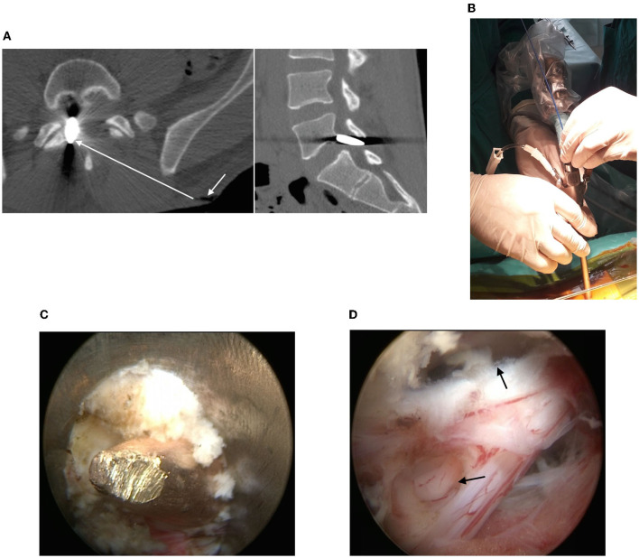
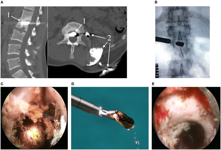
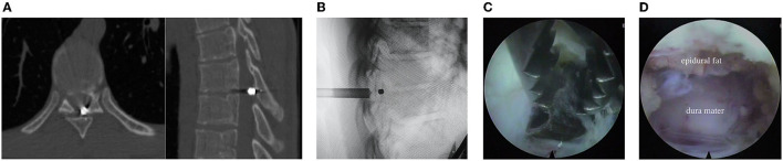
Results: Percutaneous endoscopic approach to spinal canal with a possibility to extract a bullet, decompression of nerve roots, defect closure of the dura mater is demonstrated.

Conclusion: Good clinical outcomes allows to recommend percutaneous endoscopic surgery to manage similar lumbar and thoracic spine bullet wounds at the tertiary care level.

Keywords: full-endoscopic surgery; gunshot wound; injury; lumbar spine; thoracic spine.


Conflict of interest statement

The authors declare that the research was conducted in the absence of any commercial or financial relationships that could be construed as a potential conflict of interest.

Figures

Figure 1
Figure 1
(A) CT scans of the lumbar spine: metallic foreign body (bullet) in the right half of the spinal canal (the arrow shows the inlet of the gunshot wound, the line shows an approximate trajectory of the bullet through the soft tissues); (B) View of the working port and endoscope; (C) Bullet mobilization in a yellow ligament defect; (D) Defects in the dura mater around the nerve root cuff and dural sac (see arrows).
Figure 2
Figure 2
(A) Preoperative sagittal CT (left) and CT fistulography (right) of the lumbar spine: 1—bullet; 2—contrast agent; (B) Radiography of the working port and bullet; (C) Endoscopic stage of the surgery and view of the bullet (described in the text); (D) Capturing and extracting the bullet with forceps (caliber 7.62 mm, length 15 mm); (E) Endoscopic view of the intervertebral foramen after extracting the bullet.
Figure 3
Figure 3
(A) CT scans of the thoracic spine (explained in the text); (B) Radiography of the working port and bullet; (C,D) Endoscopic stage of the surgery (described in the text).

References

    1. Shin JK, Youn MS, Seong YJ, Goh TS, Lee JS. Iatrogenic dural tear in endoscopic lumbar spinal surgery: full endoscopic dural suture repair (Youn's technique). Eur Spine J. (2018) 27:544–8. 10.1007/s00586-018-5637-6 - DOI - PubMed
    1. Telfeian AE, Veeravagu A, Oyelese AA, Gokaslan ZL. A brief history of endoscopic spine surgery. Neurosurg Focus. (2016) 40:E2. 10.3171/2015.11.FOCUS15429 - DOI - PubMed
    1. Wagner R, Telfeian AE, Iprenburg M, Krzok G, Gokaslan Z, Choi DB, et al. . Transforaminal endoscopic solution to a kyphoplasty complication. World Neurosurg. (2016) 91:195–8. 10.1016/j.wneu.2016.04.013 - DOI - PubMed
    1. Ito M, Abumi K, Kotani Y, Kadoya K, Minami A. Clinical outcome of posterolateral endoscopic surgery for pyogenic spondylodiscitis: results of 15 patients with serious comorbid conditions. Spine. (2007) 32:200–6. 10.1097/01.brs.0000251645.58076.96 - DOI - PubMed
    1. Sentürk S, Ünsal ÜÜ. Percutaneous full-endoscopic removal of lumbar intradural extramedullary tumor via translaminar approach. World Neurosurg. (2019) 125:146–9. 10.1016/j.wneu.2019.01.206 - DOI - PubMed

Publication types