

The geometric features of tibiofemoral joint associated with anterior cruciate ligament injury: Reliability of measurements on conventional MRI
- PMID: 35402659
- PMCID: PMC8989692
- DOI: 10.1016/j.ejro.2022.100420
The geometric features of tibiofemoral joint associated with anterior cruciate ligament injury: Reliability of measurements on conventional MRI
Abstract
Purpose: To evaluate the measurement reliability of the geometric features of tibiofemoral joint on conventional knee MRI and also identify the features associated with increased risk of ACL injury.
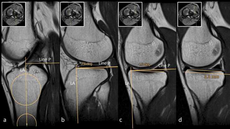
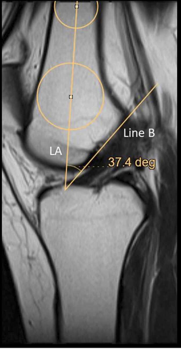
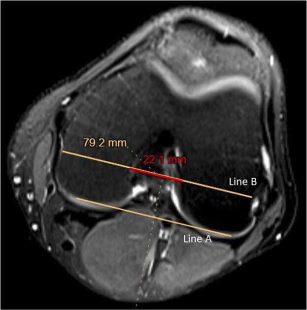
Methods: This retrospective case-control study included knee MRIs of 60 patients with ACL injury (34 men, 26 women; mean age 34 ± 13.6 SD) and 60 normal individuals (31 men, 29 women; mean age 36 ± 11.4 SD). Geometric features of distal femur (intercondylar notch width, transcondylar width, and intercondylar notch angle) and tibial plateau (medial tibial plateau slope, lateral tibial plateau slope, and medial tibial plateau depth) were independently measured by two radiologists for each of the patients. Intraclass correlation coefficient (ICC) values were calculated to assess the reliability of the measurements. Variables with acceptable ICC values were included in the final logistic regression model, but the remaining were only reported descriptively.
Results: There was good to excellent agreement between the radiologist in the measurement of ICNW and TCW. However, the agreement between the radiologists was not acceptable for the rest of the variables. The univariate logistic regression model showed as ICNW decreases, the risk of ACL injury increases (OR = 0.12, 95% CI [0.02, 0.60], p = 0.01).
Conclusions: Our results suggest that ICNW and TCW are the only geometric features of the tibiofemoral joint that can be reliably measured on conventional knee MRI. Moreover, decreased ICNW is associated with an increased risk of ACL injury.
Keywords: Anterior cruciate ligament injuries; Femur; Intercondylar notch; Reliability of results; Risk factor; Tibia; Tibial plateau.
© 2022 The Authors.
Conflict of interest statement
The authors declare that they have no known competing financial interests or personal relationships that could have appeared to influence the work reported in this paper.
Figures
References
-
- Anderson A.F., Lipscomb A.B., Liudahl K.J., Addlestone R.B. Analysis of the intercondylar notch by computed tomography. Am. J. Sports Med. 1987;15:547–552. - PubMed
-
- Ardern C.L., Webster K.E., Taylor N.F., Feller J.A. Return to sport following anterior cruciate ligament reconstruction surgery: a systematic review and meta-analysis of the state of play. Br. J. Sports Med. 2011;45:596–606. - PubMed
-
- Barenius B., Ponzer S., Shalabi A., Bujak R., Norlen L., Eriksson K. Increased risk of osteoarthritis after anterior cruciate ligament reconstruction: a 14-year follow-up study of a randomized controlled trial. Am. J. Sports Med. 2014;42:1049–1057. - PubMed
-
- Bouras T., Fennema P., Burke S., Bosman H. Stenotic intercondylar notch type is correlated with anterior cruciate ligament injury in female patients using magnetic resonance imaging. Knee Surg. Sports Trauma. Arthrosc. 2018;26:1252–1257. - PubMed
LinkOut - more resources
Full Text Sources
