. 2021 Jul 3;1(4):317-322.
doi: 10.21873/cdp.10042. eCollection 2021 Sep-Oct.

New Technique of Laparoscopic Paraaortic Lymph Node Dissection for Colorectal Cancer Using Fluorescence Navigation

Affiliations

New Technique of Laparoscopic Paraaortic Lymph Node Dissection for Colorectal Cancer Using Fluorescence Navigation

Shunjin Ryu et al. Cancer Diagn Progn. .

Abstract

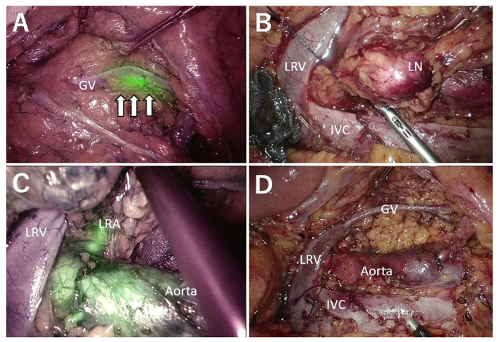
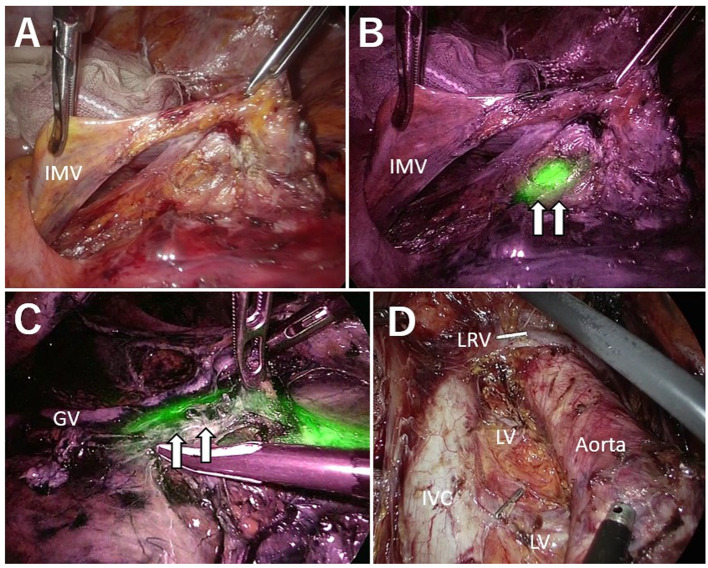
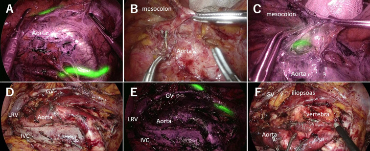
Background/aim: According to limited current reports, therapeutic paraaortic lymph node (PALN) dissection with intensive combined therapy for colorectal cancer improves prognosis in select patients. Laparoscopic PALN dissection is a difficult technique that has not yet been established. We applied this procedure using an intraoperative fluorescence navigation technique with a near-infrared ray catheter (NIRC™) fluorescent ureteral catheter (NIRFUC).

Patients and methods: We evaluated the utility of laparoscopic fluorescence navigation and the short-term outcomes of 6 patients undergoing laparoscopic PALN dissection.

Results: There were 3 surgeries for synchronous metastasis and 3 surgeries for recurrent metastasis. The mean surgical duration, blood loss, and postoperative hospital stay were 677 (range=518-1,090) min, 7.5 (range=3-1,600) ml, and 14 (range=9-33) days, respectively. Postoperative complications (Clavien-Dindo grade >III) occurred in 1 case.

Conclusion: Dissection around the ureter was navigated with a NIRFUC. Fluorescence ureteral navigation facilitated completion of the complex laparoscopic PALN dissection procedure.

Keywords: Laparoscopic surgery; colorectal cancer; fluorescence navigation; paraaortic lymph node dissection; ureter.


Conflict of interest statement

Shunjin Ryu MD, PhD, Keigo Hara MD, Keisuke Goto MD, Keisuke Goto MD, Atsuko Okamoto MD, Takahiro Kitagawa MD, Rui Marukuchi MD, PhD, Ryusuke Ito MD, PhD, and Yukio Nakabayashi MD, PhD have no conflicts of interest or financial ties to disclose in relation to this study.

Figures

Figure 1
Figure 1. Case of synchronous metastasis. A: The white arrow indicates the ureter with the NIRFUC. B: Lymph node metastasis was dissected from the LRV and IVC. Lymphatic vessels were ligated. C: FVN revealed the left renal artery with fluorescence. D: Surgical field after PALN dissection. IVC: Inferior vena cava; NIRFUC: near-infrared ray catheter (NIRC™) fluorescent ureteral catheter; FVN: fluorescence vessel navigation; PALN: paraaortic lymph node; Aorta: abdominal aorta; LRV: left renal vein; LRA: left renal artery; GV: gonadal vein.
Figure 2
Figure 2. Case of recurrence after low anterior resection. A: Under normal light observation, the ureter was not visible. On the dorsal side, the mesocolon and Gerota fascia were easily dissected. However, on the caudal side, dissection was difficult because of adhesion from a previous surgery. B: NIRFUC allowed visualization of the ureter with fluorescence. C: Lymph node dissection was performed while preserving visualization of the ureter (white arrow). D: Surgical field after paraaortic lymph node dissection. NIRFUC: near-infrared ray catheter (NIRC™) fluorescent ureteral catheter; IMV: inferior mesenteric vein; IVC: Inferior vena cava; Aorta: abdominal aorta; LRV: left renal vein; GV: gonadal vein; LV: lumbar vein.
Figure 3
Figure 3. Case of recurrence after the Hartmann procedure. A: Fluorescence observation before paraaortic lymph node (PALN) dissection. NIRFUC allowed visualization of both sides of the ureter with fluorescence. B: Dissection between the mesocolon, ureter and gonadal vein under normal light observation. The ureter was not detected. C: Fluorescence observation using the NIRFUC to visualize the ureter. D: Surgical field after PALN dissection under normal light observation. E: Surgical field after PALN dissection with fluorescence observation. F: Surgical field after PALN dissection. NIRFUC: Near-infrared ray catheter (NIRC™) fluorescent ureteral catheter; IVC: Inferior vena cava; aorta: abdominal aorta; PALN: paraaortic lymph node; LRV: left renal vein; GV: gonadal vein.

Similar articles

Cited by

References

    1. Howlader N, Noone AM, Krapcho M, Miller D, Bishop K, Kosary CL, Yu M, Ruhl J, Tatalovich Z, Mariotto A, Lewis DR, Chen HS, Feuer EJ, Cronin KA. Seer cancer statistics review, 1975-2014. Bethesda, MD, National Cancer Institute. Available at: https://seer.cancer.gov/archive/csr/1975_2014/ [Last accessed on June 14, 2021]
    1. Gagnière J, Dupré A, Chabaud S, Peyrat P, Meeus P, Rivoire M. Retroperitoneal nodal metastases from colorectal cancer: Curable metastases with radical retroperitoneal lymphadenectomy in selected patients. Eur J Surg Oncol. 2015;41(6):731–737. doi: 10.1016/j.ejso.2015.03.229. - DOI - PubMed
    1. Stearns MW Jr, Deddish MR. Five-year results of abdominopelvic lymph node dissection for carcinoma of the rectum. Dis Colon Rectum. 1959;2(2):169–172. doi: 10.1007/BF02616711. - DOI - PubMed
    1. Leggeri A, Roseano M, Balani A, Turoldo A. Lumboaortic and iliac lymphadenectomy: what is the role today. Dis Colon Rectum. 1994;37(2 Suppl):S54–S61. doi: 10.1007/BF02048433. - DOI - PubMed
    1. Yamada K, Tsukamoto S, Ochiai H, Shida D, Kanemitsu Y. Improving selection for resection of synchronous para-aortic lymph node metastases in colorectal cancer. Dig Surg. 2019;36(5):369–375. doi: 10.1159/000491100. - DOI - PubMed

LinkOut - more resources