. 2022 Aug;251(8):1306-1321.
doi: 10.1002/dvdy.475. Epub 2022 Apr 22.

Coordinated patterning of zebrafish caudal fin symmetry by a central and two peripheral organizers

Affiliations

Coordinated patterning of zebrafish caudal fin symmetry by a central and two peripheral organizers

Thomas Desvignes et al. Dev Dyn. 2022 Aug.

Abstract

Background: Caudal fin symmetry characterizes teleosts and likely contributes to their evolutionary success. However, the coordinated development and patterning of skeletal elements establishing external symmetry remains incompletely understood. We explore the spatiotemporal emergence of caudal skeletal elements in zebrafish to consider evolutionary and developmental origins of caudal fin symmetry.

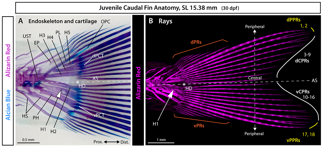
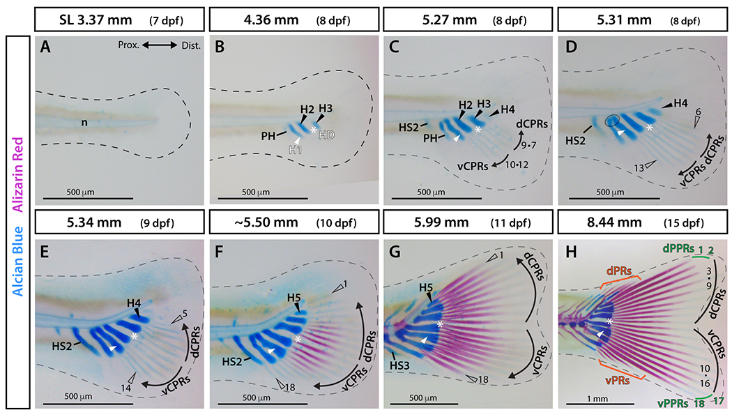
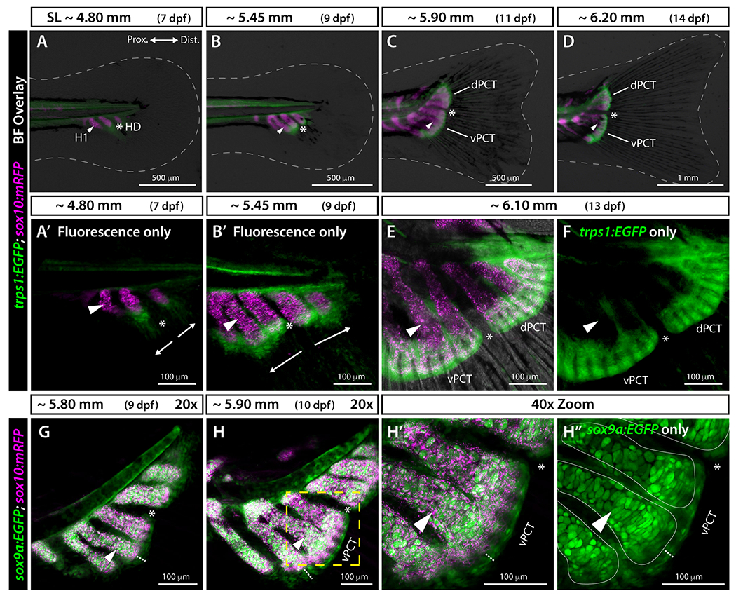
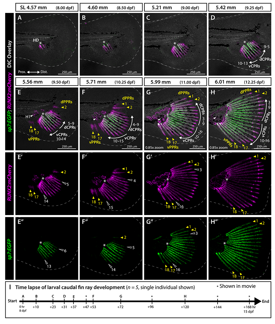
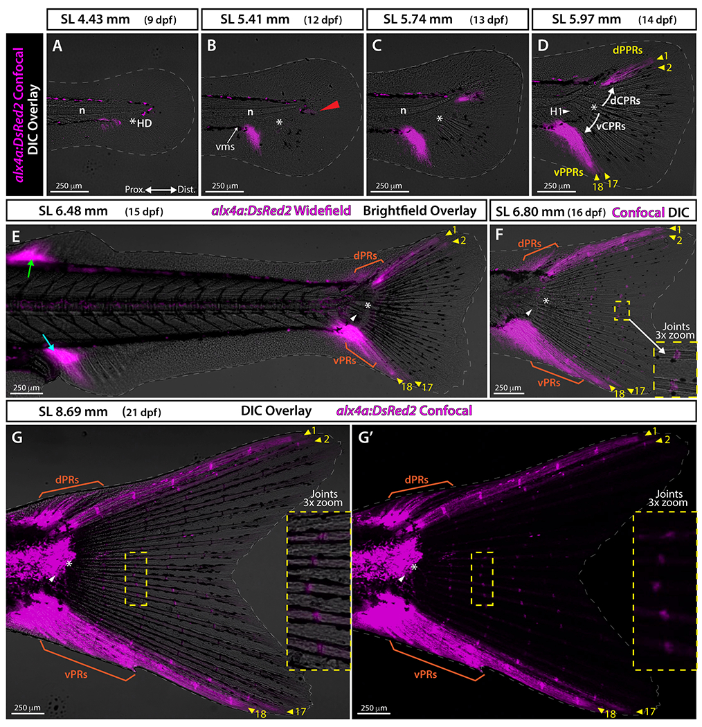
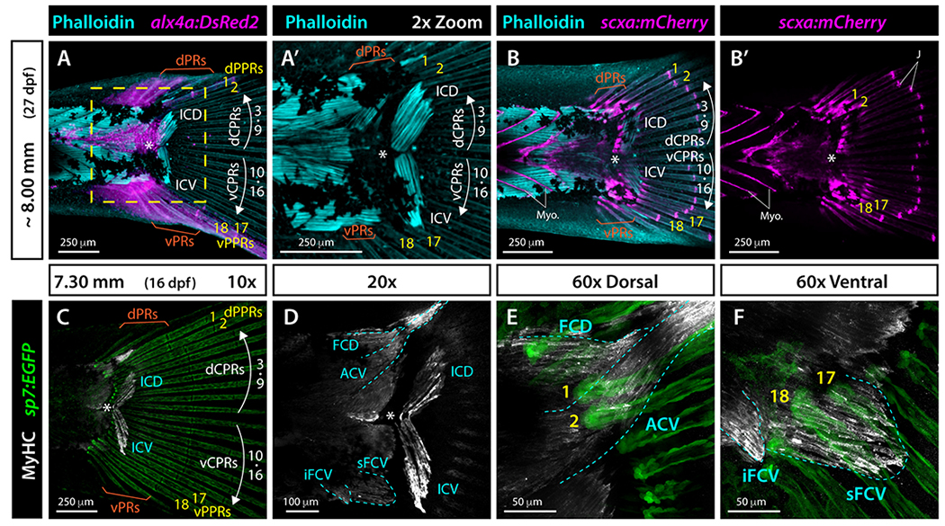
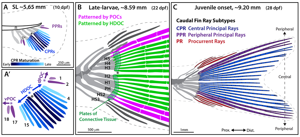
Results: Transgenic reporters and skeletal staining reveal that the hypural diastema-defining gap between hypurals 2 and 3 forms early and separates progenitors of two plates of connective tissue. Two sets of central principal rays (CPRs) synchronously, sequentially, and symmetrically emerge around the diastema. The two dorsal- and ventral-most rays (peripheral principal rays, PPRs) arise independently and earlier than adjacent CPRs. Muscle and tendon markers reveal that different muscles attach to CPR and PPR sets.

Conclusions: We propose that caudal fin symmetry originates from a central organizer that establishes the hypural diastema and bidirectionally patterns surrounding tissue into two plates of connective tissue and two mirrored sets of CPRs. Further, two peripheral organizers unidirectionally specify PPRs, forming a symmetric "composite" fin derived from three fields. Distinct CPR and PPR ontogenies may represent developmental modules conferring ray identities, muscle connections, and biomechanical properties. Our model contextualizes mechanistic studies of teleost fin morphological variation.

Keywords: actinopterygian; caudal fin; fin rays; fin symmetry; hypural diastema; teleosts.


Figures

Figure 1.
Figure 1.. Anatomy of the zebrafish caudal skeleton.
A: Whole mount brightfield and B: maximum intensity projection of confocal fluorescent images of a juvenile zebrafish caudal fin stained with Alcian Blue and Alizarin Red. The white asterisk denotes the hypural diastema (HD), the white arrowhead indicates the hypural 1 (H1), and the white dashed line marks the axis of external dorsoventral symmetry (AS). Abbreviations: dCPRs and vCPRs (white), dorsal and ventral central principal rays, respectively; dPCT and vPCT, dorsal and ventral plates of connective tissue, respectively; dPPRs and vPPRs (yellow), dorsal and ventral peripheral principal rays, respectively; dPRs and vPRs (orange), dorsal and ventral procurrent rays, respectively; EP, epural; H1-H5, hypurals 1 to 5; HS, haemal spine; OPC, opisthural cartilage; PH, parhypural; PL, pleurostyle; UST, urostyle.
Figure 2.
Figure 2.. Alcian Blue and Alizarin Red visualization of caudal fin symmetry establishment and body axis alignment anchored by the hypural diastema.
A-H: Whole-mount images of Alcian Blue (cartilage) and Alizarin Red (calcified bone) stained caudal fin skeletons across a zebrafish larval developmental sequence. The dashed line marks the border of the fin fold. The ellipse in D indicates fusion of the parhypural and hypural 1. Abbreviations: dCPRs and vCPRs, dorsal and ventral central principal rays, respectively (black); dPPRs and vPPRs, dorsal and ventral peripheral principal rays, respectively (green); dPRs and vPRs, dorsal and ventral procurrent rays, respectively (orange); H1 and white arrowhead, hypural 1; H2-5 and black arrowheads, hypurals 2 to 5; HD and white asterisk, hypural diastema; HS2-3, haemal spines 2 and 3; n, notochord; PH, parhypural.
Figure 3.
Figure 3.. Transgenic reporters reveal that plates of connective tissue develop symmetrically around the hypural diastema.
A-D: Widefield and A’, B’, E, F: confocal whole mount images of the caudal region of trps1:EGFP;sox10:mRFP larval zebrafish. White dashed line indicates fin boundaries. sox10:mRFP labels endochondral elements including hypurals, parhypural, and haemal spines, and trps1:EGFP labels precursor cells of the two plates of connective tissue. The hypural diastema lies between hypurals 2 and 3 and separates the two developing plates of connective tissue. Arrows in A’ and B’ indicate the directions of outgrowth of the plates of connective tissue. G-H”: Confocal images of sox9a:EGFP;sox10:mRFP larval zebrafish. sox9a:EGFP and sox10:mRFP transgenes are co-expressed in endochondral elements with additional sox9a:EGFP expression in the two developing plates of connective tissue separated at the hypural diastema. The white dotted lines in H-H’’ show the thickness of the plates of connective tissue. The yellow dashed line in H is blown up in H’ and H”. Solid thin white lines in H” denote the borders of the endochondral skeletal elements based on H’. Abbreviations: dPCT and vPCT, dorsal and ventral plates of connective tissue, respectively; H1 and white arrowhead, Hypural 1; HD and white asterisk, hypural diastema.
Figure 4.
Figure 4.. Osteoblast developmental state markers demonstrate synchronous and symmetric central principal ray formation and distinct emergence of peripheral principal caudal fin rays.
A-H”: Whole mount confocal images of a representative RUNX2:mCherry;sp7:EGFP larva (from n = 5) followed from SL 4.57 mm to 6.01 mm (8-12.25 dpf) and imaged following the schedule in I. Differential interference contrast (DIC) overlayed in A-H; RUNX2:mCherry (pre-osteoblasts, magenta) single channel in E’-H’; and sp7:EGFP (differentiating osteoblasts, green) single channel in E”-H”. White dashed line indicates fin boundaries. Central principal ray directions of outgrowth are indicated by white arrows in D-H. Abbreviations: dCPRs and vCPRs, dorsal and ventral central principal rays, respectively; dPPRs and vPPRs, dorsal and ventral peripheral principal rays, respectively (yellow arrowheads); H1 and white arrowhead, hypural 1; HD and white asterisk, hypural diastema. I: Timing of time lapse image acquisition points shown in the corresponding panels (letters) and additionally in Video S1 (asterisks).
Figure 5.
Figure 5.. alx4a transgenic expression indicates the distinct nature of peripheral principal rays.
A-D and F-G: Confocal images overlaid with differential interference contrast (DIC). E: Widefield fluorescence image with brightfield overlaid. Gray dashed line indicates fin boundaries. G’: alx4a:DsRed2 expression in G shown in single channel. Red arrowhead in B points at the faint alx4a:DsRed2 expression just below the notochord distal tip. White arrows in D mark the sequence of central principal ray specification. Green and cyan arrows in E point at the alx4a:DsRed2 expression at the leading rays of the dorsal and anal fins, respectively. Abbreviations: dCPRs and vCPRs, dorsal and ventral central principal rays; dPPRs and vPPRs, dorsal and ventral peripheral principal rays, respectively (yellow arrowheads); dPRs and vPRs, dorsal and ventral procurrent rays, respectively (orange); H1 and white arrowhead, Hypural 1; HD and white asterisk, hypural diastema; n, notochord; vms, ventral melanophore stripe before the caudal fin condensation region.
Figure 6.
Figure 6.. Muscle staining combined with alx4a and scxa reporters reveal that each set of central principal rays and peripheral principal rays is associated with distinct swimming muscles.
Whole mount widefield fluorescent images of the caudal region of zebrafish larvae. A, A’: Phalloidin-stained ~8.00 mm alx4a:DsRed2 larval zebrafish. F-actin (phalloidin) is cyan and alx4a:DsRed2-expressing cells, including peripheral principal rays (PPRs), are magenta. B-B’: Phalloidin-stained (cyan) ~8.00 mm scxa:mCherry larval zebrafish. The mCherry fluorescent marker (magenta) labels tendons, myosepta, and caudal fin ray joints. C-F: Whole mount immunostained 7.30 mm sp7:EGFP (green) larva. Myosin heavy chain (Myh) antibody labels skeletal muscles (white). EGFP fluorescence is green. Muscles are outlines in cyan dashed line. Abbreviations: ACV, abductor caudalis ventralis; dCPR and vCPR, dorsal and ventral central principal rays, respectively; dPPR and vPPR (yellow), dorsal and ventral peripheral principal rays, respectively; dPR and vPR, dorsal and ventral procurrent rays, respectively; FCD, flexor caudalis dorsalis; ICD, inerfilamenti caudalis dorsalis; ICV, inerfilamenti caudalis ventralis; iFCV, inferior flexor caudalis ventralis; J, ray joints; Myo, myosepta; sFCV, superior flexor caudalis ventralis; white asterisk, hypural diastema.
Figure 7.
Figure 7.. Schematic representation of zebrafish caudal fin skeletal patterning.
The caudal fin comprises three subtypes of fin rays differing in their ontogeny, morphology, and identity: central principal rays (CPRs), two sets of peripheral principal rays (ventral and dorsal PPRs), and procurrent rays (PRs). A-A’: CPRs (blue, with dark-to-light color gradient representing the first-to-last forming rays) are specified by a hypothetical bidirectional hypural diastema organizing center (HDOC; blue sphere) triggering the sequential and synchronous addition of mirrored pairs of rays (14 total) symmetrically distributed around the hypural diastema (asterisk). Two sets of PPRs (principal rays 1, 2 and 17, 18; purple) develop separately from the CPRs, unidirectionally specified by two hypothetical peripheral organizing centers (POCs, purple spheres). (B) Two plates of connective tissue (light green) are specified on each side of the HDOC along with CPRs (green), while PPRs (pink) are specified by POCs. H1-H5, Hypurals 1-5; HS2 and HS3, haemal spines 2 and 3. (C) CPRs (blue) are segmented and branched. Among PPRs (purple), the two most peripheral (principal rays 1 and 18) are segmented but unbranched, while PPR 2 and 17 are segmented and branched. Unbranched PRs (red) emerge after the formation of all principal rays. This model proposes that the caudal fin is a “compound” appendage with body axis-aligned external symmetry arising from 1) HDOC-initiated, symmetrically specified CPRs and plates of connective tissue, 2) two similar PPR sets specified by two POCs equally spaced from the HDOC, and 3) upward bending of the notochord that rotates skeletal elements from an anterior-posterior to a dorsal-ventral position, aligning the hypural diastema with the body axis.

References

    1. Giammona FF. Form and Function of the Caudal Fin Throughout the Phylogeny of Fishes. Integrative and Comparative Biology. 2021;61(2):550–572. doi:10.1093/icb/icab127 - DOI - PubMed
    1. Gosline WA. Functional morphology of the caudal skeleton in teleostean fishes. Ichthyological Research. 1997;44(2–3):137–141. doi:10.1007/BF02678693 - DOI
    1. Lauder GV. Function of the Caudal Fin During Locomotion in Fishes: Kinematics, Flow Visualization, and Evolutionary Patterns. American Zoologist. 2000;40(1):101–122. doi:10.1668/0003-1569(2000)040[0101:FOTCFD]2.0.CO;2 - DOI
    1. Schultze HP, Arratia G. The composition of the caudal skeleton of teleosts (Actinopterygil: Osteichthyes). Zoological Journal of the Linnean Society. 1989;97(3):189–231. doi:10.1111/j.1096-3642.1989.tb00547.x - DOI
    1. Bensimon-Brito A, Cancela ML, Huysseune A, Witten PE. Vestiges, rudiments and fusion events: the zebrafish caudal fin endoskeleton in an evo-devo perspective. Evol Dev. 2012;14(1):116–127. doi:10.1111/j.1525-142X.2011.00526.x - DOI - PubMed

Publication types

LinkOut - more resources