Review
. 2022 Apr 15;23(4):265-285.
doi: 10.1631/jzus.B2100977.

Molecular pathogenesis of acetaminophen-induced liver injury and its treatment options

Affiliations
Review

Molecular pathogenesis of acetaminophen-induced liver injury and its treatment options

Xiaopeng Cai et al. J Zhejiang Univ Sci B. .

Abstract

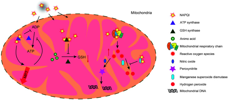
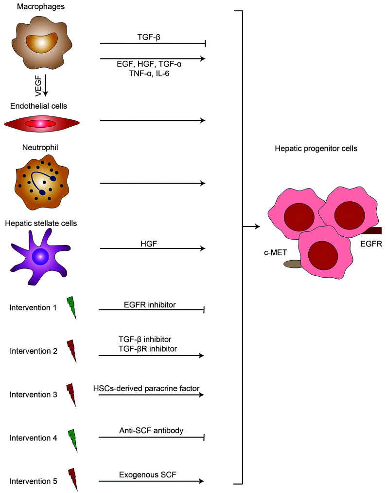
Acetaminophen, also known as N-acetyl-p-aminophenol (APAP), is commonly used as an antipyretic and analgesic agent. APAP overdose can induce hepatic toxicity, known as acetaminophen-induced liver injury (AILI). However, therapeutic doses of APAP can also induce AILI in patients with excessive alcohol intake or who are fasting. Hence, there is a need to understand the potential pathological mechanisms underlying AILI. In this review, we summarize three main mechanisms involved in the pathogenesis of AILI: hepatocyte necrosis, sterile inflammation, and hepatocyte regeneration. The relevant factors are elucidated and discussed. For instance, N-acetyl-p-benzoquinone imine (NAPQI) protein adducts trigger mitochondrial oxidative/nitrosative stress during hepatocyte necrosis, danger-associated molecular patterns (DAMPs) are released to elicit sterile inflammation, and certain growth factors contribute to liver regeneration. Finally, we describe the current potential treatment options for AILI patients and promising novel strategies available to researchers and pharmacists. This review provides a clearer understanding of AILI-related mechanisms to guide drug screening and selection for the clinical treatment of AILI patients in the future.

Keywords: Acetaminophen; Acetaminophen-induced liver injury; Hepatocyte necrosis; Hepatocyte regeneration; Sterile inflammation.


Figures

Fig. 1
Fig. 1. Schematic diagram of APAP metabolism. Under the action of glucoronosyl-transferase and sulfo-transferase, most monomer APAP forms complexes with sulfate and glucuronic acid, which are excreted in bile or urine. The remaining part of APAP is oxidized by CYP450 to form NAPQI, which combines with GSH. Accompanied with overconsumption of GSH, NAPQI shows accumulation in the liver, eventually causing liver damage. APAP: N-acetyl-p-aminophenol; CYP450: cytochrome P450; NAPQI: N-acetyl-p-benzoquinone imine; GSH: glutathione.
Fig. 2
Fig. 2. Mitochondrial oxidative/nitrosative stress and dysfunction. Excessive NAPQI in the mitochondria combines with ATP synthase, GSH synthase, and oxidative respiratory chain enzymes to form protein adducts, which subsequently lead to mitochondrial dysfunction and oxidative stress. NAPQI-ATP synthase hinders ATP synthesis and gradually triggers the opening of MPT pores. The abnormality of MPT pores is exacerbated by the inhibition of ATP synthesis. NAPQI-GSH synthase reduces GSH synthesis, weakens the excretion of NAPQI, and induces the formation of ROS. ROS can be eliminated by the antioxidant enzyme system, but excessive ROS combine with free NO to form peroxynitrite, causing mitochondrial DNA damage. NAPQI: N-acetyl-p-benzoquinone imine; ADP: adenosine diphosphate; ATP: adenosine triphosphate; GSH: glutathione; ROS: reactive oxygen species; MPT: membrane permeability transition; NO: nitric oxide.
Fig. 3
Fig. 3. Roles of JNK, P38, and ERK activation in AILI. NAPQI in hepatocytes binds to enzymes on the MRC to produce ROS, which are released into the matrix, and phosphorylates JNK through three different pathways. Phosphorylated JNK (p-JNK) is ectopic to the vicinity of mitochondria, and combines with Sab to affect the electron transmission of MRC and induce ROS production, indicating the existence of a feedback loop between ROS and JNK. Persistent mitochondrial oxidative stress and dysfunction contribute to the opening of MPT pores and leakage of mitochondrial solute into the matrix, such as AIF, endonuclease (Endo) G, and cytochrome C. This induces nucleic acid cleavage and necrosis in liver cells. The stress response of the ER aggravates liver cell death. Hepatocytes produce self-defense reactions to deal with the damage, including the production of P53 protein and autophagy. P53 protein reduces mitochondrial damage by inhibiting p-JNK. Autophagosomes can clear up the protein adducts and the damaged mitochondria, to regulate ER transition. P38 and ERK are activated in the process of AILI. The activation of ERK induces hepatocyte apoptosis and pro-inflammatory gene expression of inflammatory cells. JNK: c-Jun N-terminal kinase; ERK: extracellular signal-regulated kinase; AILI: acetaminophen-induced liver injury; NAPQI: N-acetyl-p-benzoquinone imine; MRC: mitochondrial respiratory chain; ROS: reactive oxygen species; MPT: membrane permeability transition; AIF: apoptosis-inducing factor; Sab: SH3 homology-associated bruton tyrosine kinase-binding protein; P: phosphorylated; GSK-3β: glycogen synthase kinase 3β; MLK3: mixed lineage kinase 3; MKK: mitogen-activated protein kinase kinase; ASK: apoptosis signaling-regulating kinase; ULK: unc-51-like kinase; GSH: glutathione; ER: endoplasmic reticulum; CHOP: CCAAT/enhancer-binding protein homologous protein.
Fig. 4
Fig. 4. Involvement of sterile inflammation in AILI. Liver KCs are activated by different types of DAMPs. Then KCs affect the life cycle of hepatocytes by releasing different inflammatory mediators, and/or induce phenotypic changes in other immune cells. Neutrophils serve as scavengers without aggravating liver damage, and they can promote liver cell recovery and the maturation of MoMFs. MoMFs show crosstalk with neutrophils. Ly-6Chigh MoMF can promote neutrophil chemotaxis, but Ly-6Clow MoMF inhibits neutrophil recruitment and participates in tissue repair. While the expression of MHC-I molecules decreases in necrotic liver cells, NK/NKT cells are activated and the toxic hepatocytes are scavenged. The gut-liver axis has an important function during AILI, and intestinal dysbiosis will aggravate AILI. AILI: acetaminophen-induced liver injury; KCs: Kupffer cells; DAMP: damage-related molecular pattern; MoMF: monocyte-derived macrophage; Ly-6C: lymphocyte antigen 6C; MHC-I: major histocompatibility complex-I; NK: nature killer; NKT: natural killer T; IL: interleukin; MIP1/2: macrophage inflammatory protein 1/2; RAGE: receptor for advanced glycation end products; TLR: Toll-like receptor; MCP-1: monocyte chemoattractant protein-1; ROS: reactive oxygen species; HMGB1: high mobility group box 1 protein; TNF-α: tumor necrosis factor-α; IFN-γ: interferon-γ; GSH: glutathione; CYPE1: cytochrome P450 E1.
Fig. 5
Fig. 5. HPCs regenerated under the action of signal mediators and immune cells. Immune cells (e.g., macrophages and neutrophils) involved in the scavenging of necrotic liver cells and tissues provide space for the survival of HPCs. Meanwhile, the vascular endothelium proliferates under the action of VEGF and participates in liver microangiogenesis. The nonparenchymal cells of the liver can secrete various cytokines, for example, EGF and HGF, and promote the regeneration of hepatic progenitor cells through different signal proliferation pathways. The regeneration process of hepatocytes can be affected through external intervention, for instance, the use of EGFR inhibitors, TGF inhibitors, or HSCs. HPCs: hepatic progenitor cells; TGF: transforming growth factor; TGF-βR: TGF-β receptor; TNF-α: tumor necrosis factor-α; VEGF: vascular endothelial growth factor; EGF: epidermal growth factor; HGF: hepatocytes growth factor; IL: interleukin; EGFR: EGF receptor; HSCs: hepatic stellate cells; SCF: stem cell factor; c-MET: cellular mesenchymal epithelial transition factor.

References

    1. Akakpo JY, Ramachandran A, Kandel SE, et al. , 2018. 4-Methylpyrazole protects against acetaminophen hepatotoxicity in mice and in primary human hepatocytes. Hum Exp Toxicol, 37(12): 1310-1322. 10.1177/0960327118774902 - DOI - PMC - PubMed
    1. Akakpo JY, Ramachandran A, Jaeschke H, 2020. Novel strategies for the treatment of acetaminophen hepatotoxicity. Expert Opin Drug Metab Toxicol, 16(11): 1039-1050. 10.1080/17425255.2020.1817896 - DOI - PMC - PubMed
    1. Antoniades CG, Quaglia A, Taams LS, et al. , 2012. Source and characterization of hepatic macrophages in acetaminophen-induced acute liver failure in humans. Hepatology, 56(2): 735-746. 10.1002/hep.25657 - DOI - PubMed
    1. Apte U, Singh S, Zeng G, et al. , 2009. Beta-catenin activation promotes liver regeneration after acetaminophen-induced injury. Am J Pathol, 175(3): 1056-1065. 10.2353/ajpath.2009.080976 - DOI - PMC - PubMed
    1. Barbier-Torres L, Iruzubieta P, Fernández-Ramos D, et al. , 2017. The mitochondrial negative regulator MCJ is a therapeutic target for acetaminophen-induced liver injury. Nat Commun, 8: 2068. 10.1038/s41467-017-01970-x - DOI - PMC - PubMed

MeSH terms