

3D imaging for driving cancer discovery
- PMID: 35403737
- PMCID: PMC9108604
- DOI: 10.15252/embj.2021109675
3D imaging for driving cancer discovery
Abstract
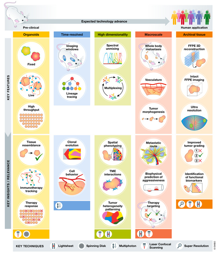
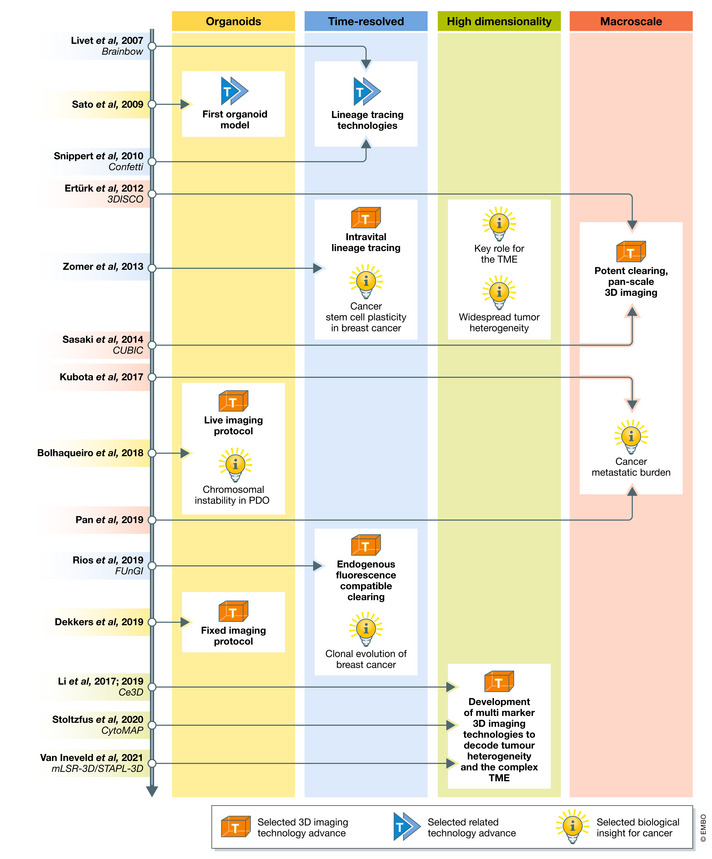
Our understanding of the cellular composition and architecture of cancer has primarily advanced using 2D models and thin slice samples. This has granted spatial information on fundamental cancer biology and treatment response. However, tissues contain a variety of interconnected cells with different functional states and shapes, and this complex organization is impossible to capture in a single plane. Furthermore, tumours have been shown to be highly heterogenous, requiring large-scale spatial analysis to reliably profile their cellular and structural composition. Volumetric imaging permits the visualization of intact biological samples, thereby revealing the spatio-phenotypic and dynamic traits of cancer. This review focuses on new insights into cancer biology uniquely brought to light by 3D imaging and concomitant progress in cancer modelling and quantitative analysis. 3D imaging has the potential to generate broad knowledge advance from major mechanisms of tumour progression to new strategies for cancer treatment and patient diagnosis. We discuss the expected future contributions of the newest imaging trends towards these goals and the challenges faced for reaching their full application in cancer research.
Keywords: 3D imaging; archival tissue; cancer (immuno)therapy; cancer biology.
© 2022 The Authors.
Figures
References
-
- (2018) Tumor cells metastasize from lymph nodes. Cancer Discov 8: OF10 - PubMed
Publication types
MeSH terms
LinkOut - more resources
Full Text Sources
Medical
