. 2022 May 11;96(9):e0213721.
doi: 10.1128/jvi.02137-21. Epub 2022 Apr 11.

UV Irradiation of Vaccinia Virus-Infected Cells Impairs Cellular Functions, Introduces Lesions into the Viral Genome, and Uncovers Repair Capabilities for the Viral Replication Machinery

Affiliations

UV Irradiation of Vaccinia Virus-Infected Cells Impairs Cellular Functions, Introduces Lesions into the Viral Genome, and Uncovers Repair Capabilities for the Viral Replication Machinery

Conor W Templeton et al. J Virol. .

Abstract

Vaccinia virus (VV), the prototypic poxvirus, encodes a repertoire of proteins responsible for the metabolism of its large dsDNA genome. Previous work has furthered our understanding of how poxviruses replicate and recombine their genomes, but little is known about whether the poxvirus genome undergoes DNA repair. Our studies here are aimed at understanding how VV responds to exogenous DNA damage introduced by UV irradiation. Irradiation of cells prior to infection decreased protein synthesis and led to an ∼12-fold reduction in viral yield. On top of these cell-specific insults, irradiation of VV infections at 4 h postinfection (hpi) introduced both cyclobutene pyrimidine dimer (CPD) and 6,4-photoproduct (6,4-PP) lesions into the viral genome led to a nearly complete halt to further DNA synthesis and to a further reduction in viral yield (∼35-fold). DNA lesions persisted throughout infection and were indeed present in the genomes encapsidated into nascent virions. Depletion of several cellular proteins that mediate nucleotide excision repair (XP-A, -F, and -G) did not render viral infections hypersensitive to UV. We next investigated whether viral proteins were involved in combatting DNA damage. Infections performed with a virus lacking the A50 DNA ligase were moderately hypersensitive to UV irradiation (∼3-fold). More strikingly, when the DNA polymerase inhibitor cytosine arabinoside (araC) was added to wild-type infections at the time of UV irradiation (4 hpi), an even greater hypersensitivity to UV irradiation was seen (∼11-fold). Virions produced under the latter condition contained elevated levels of CPD adducts, strongly suggesting that the viral polymerase contributes to the repair of UV lesions introduced into the viral genome. IMPORTANCE Poxviruses remain of significant interest because of their continuing clinical relevance, their utility for the development of vaccines and oncolytic therapies, and their illustration of fundamental principles of viral replication and virus/cell interactions. These viruses are unique in that they replicate exclusively in the cytoplasm of infected mammalian cells, providing novel challenges for DNA viruses. How poxviruses replicate, recombine, and possibly repair their genomes is still only partially understood. Using UV irradiation as a form of exogenous DNA damage, we have examined how vaccinia virus metabolizes its genome following insult. We show that even UV irradiation of cells prior to infection diminishes viral yield, while UV irradiation during infection damages the genome, causes a halt in DNA accumulation, and reduces the viral yield more severely. Furthermore, we show that viral proteins, but not the cellular machinery, contribute to a partial repair of the viral genome following UV irradiation.

Keywords: DNA damage; DNA polymerase; DNA repair; DNA replication; UV irradiation; poxvirus; vaccinia.


Conflict of interest statement

The authors declare no conflict of interest.

Figures
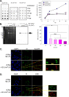
FIG 1
FIG 1
UV irradiation during VV infection introduces UV lesions into the VV genomes and halts further VV DNA accumulation. UV irradiation (30 or 60 J/m2) at 4 hpi halts further viral DNA accumulation (A and B) and introduces UV lesions, both 6,4-photoproducts (6,4-PP) and cyclobutane pyrimidine dimers (CPD), into the viral genome (C and D). (A) BSC40 cells were either left uninfected or infected with WT virus (MOI of 5) and left unirradiated (blue) or irradiated at 4 hpi (30 or 60 J/m2, pink or purple, respectively). Samples were harvested at 3, 4, 4.5, 7, or 10 hpi, and the levels of viral DNA were assessed by Southern dot blot analysis. Each sample was spotted in technical triplicate (left panel); a representative blot is shown. The data are plotted as the average of three biological experiments with the error bars representing the standard errors of the mean (SEM; right panel; ***, P < 0.001). (B) BSC40 cells were infected with WT virus (MOI of 5) and either left unirradiated or UV irradiated (15, 30, 45, or 60 J/m2) at 4 hpi before being collected at 10 hpi and processed for PFGE analysis. A representative image of the EtBr staining is shown (left), as well as the corresponding Southern blot (right); the arrow marks the monomeric viral genome. The data are plotted as the averages of six biological replicates with the error bars representing the SEM (right panel; ***, P < 0.001). (C and D) BSC40 cells were infected with WT virus (MOI of 5) for 4 h and left unirradiated or UV irradiated with 60 J/m2 before being fixed at 4.5 hpi and stained with DAPI (blue), anti-I3 (green), and either anti-6,4-PP (C; red) or anti-CPD (D; red). Both 6,4-PPs and CPDs localize to the viral cytoplasmic replication factories following UV irradiation at 4 hpi. Scale bar, 100 μm.
FIG 2
FIG 2
UV irradiation during infection reduces viral infectious yield and late protein accumulation in a dose-dependent manner. Late protein accumulation and viral infectious yield are reduced in a dose-dependent manner following UV irradiation at 4 hpi. BSC40 cells were infected with WT virus (MOI of 5). At 4 hpi, samples were either left unirradiated (blue) or irradiated with 15, 30, 45, or 60 J/m2 (pink to purple). All samples were harvested at 18 hpi and either processed for immunoblotting (A) or titrated via plaque assay to assess viral yield (B). Immunoblots were probed for calnexin (loading control), p53, I3, L4, and F17 (viral proteins) (n = 3; a representative immunoblot is shown). Viral yield is plotted as the average of three biological replicates with the error bars representing the SEM (**, P < 0.01; ***, P < 0.001).
FIG 3
FIG 3
UV irradiation prior to infection reduces viral infectious yield but not DNA accumulation. Although UV irradiation prior to infection reduces viral infectious yield, protein synthesis and late protein accumulation, viral DNA accumulation is unaffected. (A) UV irradiation at −1 hpi reduces viral yield. BSC40 cells were infected with WT virus (MOI of 5) for 18 h and were either left unirradiated or UV irradiated 1 h prior to infection (–1 h) or at 4 hpi (4 h) with either 30 or 60 J/m2. The viral yield was assessed via plaque assay (n = 3, average and SEM plotted; **, P < 0.01). (B) Viral infection at 4 hpi, but not at −1 hpi, reduces DNA accumulation. Following the same experimental design used for panel A, samples were collected at 10 hpi, and the levels of viral DNA assessed by Southern dot blotting (n = 3, average and SEM plotted; ***, P < 0.001). (C) UV irradiation at both −1 hpi and 4 hpi reduces late protein accumulation. The samples described in panel A were also subjected to immunoblot analysis. Blots were probed for calnexin, I3, L4, and F17 (n = 3, a representative immunoblot is shown). (D) UV irradiation at both −1 hpi or 4 hpi reduces protein synthesis. BSC40 cells were either uninfected (lanes 1 and 2) or infected with WT virus (MOI of 5) (lanes 3 to 14); cells were also either left unirradiated (lanes 1 and 3 to 6) or irradiated at −1 hpi (lanes 2 and 7 to 10) or 4 hpi (lanes 11 to 14). At each time point (uninfected or 3, 6, 9, and 12 hpi), the cells were labeled with [35S]methionine for 45 min. The cell lysates were resolved by SDS-PAGE to visualize the profile of nascent proteins (n = 3; a representative image is shown). (E) UV irradiation results in increased levels of phosphorylated initiation factor eIF2α in both uninfected and infected cells. BSC40 cells were either left uninfected (lanes 4 to 6, 10, and 11) or infected with WT virus (MOI of 5) (lanes 1 to 3, 7 to 9, 12, and 13); cells were also either left unirradiated (lanes 1 to 3) or irradiated at −1 hpi (lanes 4 to 9) or 4 hpi (lanes 10 to 14). At each time point (3, 6, or 9 hpi), cells were collected and processed for immunoblot analysis. Blots were probed for ATR, calnexin (loading control), Ku70, pChk1 S345, tChk1, eIF2α, p-eIF2α, F17, and L2 (viral proteins) (n = 3, a representative immunoblot is shown).
FIG 4
FIG 4
UV irradiation prior to or during infection causes defects in virion maturation. UV irradiation has a modest impact on the profile of virion assembly. BSC40 cells were either left unirradiated (top) or irradiated prior to (−1 hpi, bottom) or after (4 hpi, middle) infection. Cells were infected with WT virus (MOI of 5) for 18 h before being processed for electron microscopy. Representative images are shown. Labels: C, crescent membranes; IV, immature virions; IVN, immature virions with nucleoids; MV, mature virions; *, aberrant virion. Scale bars, 200 nm.
FIG 5
FIG 5
UV irradiation prior to or during infection reduces both virion number and infectivity. UV irradiation prior to or during infection reduces the yield of mature virions. BSC40 cells were infected with WT virus (MOI of 5) and either left unirradiated or UV irradiated prior to (−1 h) or after (4 h) infection. At 18 hpi, samples were collected, and virions were purified on sucrose gradients. Light scattering bands were photographed (A) before fractions were dripped and collected for analysis by Southern dot blot (B), immunoblot (C), plaque assay (D), and PFGE (E). (B) Virions produced from infections UV irradiated prior to or postinfection contain less DNA than WT virions. Volumes for Southern dot blot were adjusted to load equal amounts of virions based on the intensity of the light scattering band and spotted in technical duplicate. (C) Encapsidated viral proteins are reduced in UV irradiated infections. Peak fractions identified by BCA were immunoblotted for L4 and F17. (D) The infectious yield produced following UV irradiation prior to or postinfection is also reduced compared to WT virions; virions produced from cells irradiated at 4 hpi contain CPD lesions. Fractions 11 through 14 were combined and titrated on BSC40 cells (n = 1; t = 3, average and SEM plotted; *, P < 0.05). (E) UV irradiation during infection results in the encapsidation of viral genomes containing UV lesions. The pooled fractions were also resolved in duplicate by PFGE and assessed by EtBr staining and Southern blotting with a 32P-labeled viral DNA probe or with the CPD recognizing antibody.
FIG 6
FIG 6
UV lesions are long-lived in VV genomes. When infected cells are irradiated at 4 hpi, a 2-fold increase in monomeric genomes and a corresponding decrease in CPD lesions is observed at late times of infection (10 to 18 hpi). BSC40 cells were left uninfected or infected with WT virus (MOI of 5) and either left unirradiated or irradiated with 60 J/m2 UV at 4 hpi. (A) Samples were collected at 4.5, 7, 10, and 18 hpi and processed for PFGE analysis. DNA transferred to nitrocellulose filters was probed with either 32P-labeled viral DNA (left) or anti-CPD antibody (right) (n = 4, a representative image is shown). (B) Data were quantified, normalized, and plotted as the percentage of the highest value for each curve (average of n = 4). (C) BSC40 cells were infected with WT virus (MOI of 5) and left unirradiated or irradiated with 60 J/m2 UV at 4 hpi. Samples were collected at 4.5, 10, and 18 hpi and processed for PFGE analysis. Southern blots were performed with 32P-labeled viral DNA, and the signal for monomeric viral genomes is plotted. Error bars represent the SEM.
FIG 7
FIG 7
Depletion of XP-A, -F, or -G does not render infections hypersensitive to UV irradiation. (A) Generation of BSC40 cells with stable depletion of XP proteins. BSC40 cells were stably depleted of either XP-A, -F, or -G by lentiviral delivery of shRNA and selection with Puro; immunoblot analysis confirmed knockdowns of 85 to 95% (n = 3, a representative image is shown). Multiple shRNA sequences were tested and the sequence that gave the greatest depletion (underlined) was used for experimentation. (B) Depletion of XP-A, -F, and -G do not lead to UV hypersensitivity of viral infections. Transduced BSC40 cells were infected with WT virus (MOI of 5) and left unirradiated or irradiated with 60 J/m2 UV at 4 hpi. At 10 hpi, samples were collected and processed for Southern dot blot analysis (B, column 2). At 18 hpi, samples were collected and titrated on BSC40 cells to quantify viral infectious yield (B, column 1) (n = 3; *, P < 0.05).
FIG 8
FIG 8
Viruses lacking the A50 DNA ligase gene are hypersensitive to UV irradiation. (A) Viruses lacking the A50 gene are hypersensitive to UV irradiation at 4 hpi. BSC40 cells were left unirradiated (lanes 1 and 4) or irradiated with 60 J/m2 UV at 1 h prior to infection (lanes 2 and 5) or 4 hpi (lanes 3 and 6). Infections were performed with either WT virus (lanes 1 to 3) or a virus lacking A50 (vΔA50, lanes 4 to 6). Samples were collected at 18 hpi and analyzed by plaque assay (A, n = 3, average and SEM plotted; *, P < 0.05). or immunoblot (B, n = 3, representative immunoblot shown). (C and D) Viral DNA accumulation after UV irradiation is halted when viruses lack the A50 gene. BSC40 cells were left uninfected (lane 1), infected with WT virus (lanes 2, 3, and 5), or vΔA50 (lanes 4 and 6) virus. At 4 hpi, infected cultures were irradiated with 60 J/m2 UV (lanes 5 and 6). At 18 hpi, samples were collected, resolved by PFGE, stained with EtBr (8C, left), and analyzed by Southern blotting using a 32P-labeled viral DNA probe (panel C, right). (D) Under the same conditions described for panel C, cultures were collected at 10, 14, and 18 hpi before analyzing via Southern dot blotting. Data were quantified and plotted as the average of three biological replicates (error bars = the SEM; *, P < 0.05; **, P < 0.01).
FIG 9
FIG 9
Inhibition of the viral DNA polymerase at the time of UV irradiation leads to a significant further reduction in the production of infectious virus. Inhibition of the E9 polymerase by araC greatly increases the sensitivity of VV infection when UV irradiation is performed at 4 hpi, but not at −1 hpi. (A) Irradiation prior to infection: BSC40 cells were left unirradiated (lanes 1, 2, and 3) or irradiated with 60 J/m2 UV at −1 hpi (lanes 4 and 5). Cells were then infected with WT virus (MOI of 5). araC (20 μM) was added to infections 30 min (lane 2) or 4 h (lanes 3 and 5) after infection. At 18 h, samples were collected and analyzed by plaque assay (top) or immunoblot (bottom). (B) Irradiation during infection: the same experiment as described above was performed, but UV irradiation was performed at 4 hpi (n = 3, average and SEM plotted [A]; *, P < 0.05; **, P < 0.01; ***, P < 0.001, or a representative immunoblot shown [B]).
FIG 10
FIG 10
Inhibition of the E9 polymerase’s activity after UV irradiation leads to the production of virions that have a lower infectivity and contain genomes with a higher level of CPD lesions. UV irradiation at 4 hpi (with or without the addition of araC) at the time of irradiation reduces mature virion production. BSC40 cells were infected with WT virus (MOI of 5) and left unirradiated (sample 1) or irradiated at 4 hpi (samples 2 and 3); araC (20 μM) was added to sample 3 immediately thereafter. Samples were collected at 18 hpi and virions were purified; light scattering bands were photographed (A) before fractions were collected for analysis by immunoblot (B), plaque assay (C), and PFGE (D). (B) Virions purified from cells that were UV irradiated and immediately treated with araC contain lower levels of some core proteins (L4 and I6). Blots were probed for L4, F17, I6, and A30. (C) The infectivity of virions produced following UV irradiation and araC treatment is greatly reduced. Fractions 8 to 11 were pooled and titrated on BSC40 cells. (D) The genomes encapsidated within virions produced following UV irradiation and araC treatment contain an elevated level of CPD lesions. Fractions 8 to 11 were pooled, resolved by PFGE and analyzed by EtBr staining and hybridization with a 32P-labeled viral DNA fragment or by probing with an anti-CPD antibody. (The upper portion of the blot in the anti-CPD panel represents monomeric viral genomes and was exposed for a longer time than the lower level of the blot, which corresponds to fragmented cellular DNA [n = 1, t = 3, average and SEM plotted; **, P < 0.01; ***, P < 0.001].)

References

    1. Moss B. 2013. Poxviridae, p 2129–2159. In Knipe DM, Howley PM (ed), Fields virology. Lippincott/Williams & Wilkins, New York, NY.
    1. Moss B. 2013. Poxvirus DNA replication. Cold Spring Harb Perspect Biol 5:a010199. - PMC - PubMed
    1. Challberg MD, Englund PT. 1979. Purification and properties of the deoxyribonucleic acid polymerase induced by vaccinia virus. J Biol Chem 254:7812–7819. 10.1016/S0021-9258(18)36019-8. - DOI - PubMed
    1. McDonald WF, Traktman P. 1994. Vaccinia virus DNA polymerase: in vitro analysis of parameters affecting processivity. J Biol Chem 269:31190–31197. 10.1016/S0021-9258(18)47408-X. - DOI - PubMed
    1. McDonald WF, Klemperer N, Traktman P. 1997. Characterization of a processive form of the vaccinia virus DNA polymerase. Virology 234:168–175. 10.1006/viro.1997.8639. - DOI - PubMed