. 2022 Apr 27;10(2):e0019522.
doi: 10.1128/spectrum.00195-22. Epub 2022 Apr 11.

Genome-Wide Investigation of Pasteurella multocida Identifies the Stringent Response as a Negative Regulator of Hyaluronic Acid Capsule Production

Affiliations

Genome-Wide Investigation of Pasteurella multocida Identifies the Stringent Response as a Negative Regulator of Hyaluronic Acid Capsule Production

Thomas R Smallman et al. Microbiol Spectr. .

Abstract

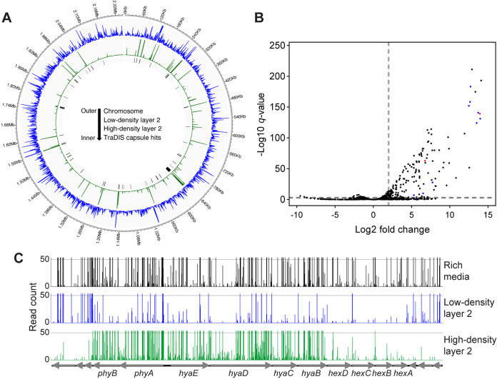
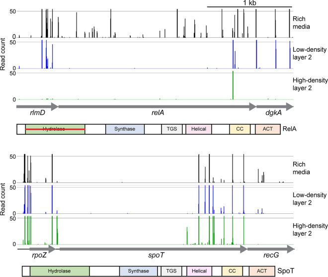
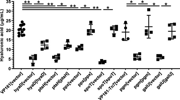
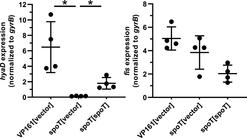
Pasteurella multocida is a Gram-negative capsulated bacterium responsible for a range of diseases that cause severe morbidity and mortality in livestock animals. The hyaluronic acid (HA) capsule produced by P. multocida serogroup A strains is a critical virulence factor. In this study, we utilized transposon-directed insertion site sequencing (TraDIS) to identify genes essential for in vitro growth of P. multocida and combined TraDIS with discontinuous density gradients (TraDISort) to identify genes required for HA capsule production and regulation in this pathogen. Analysis of mutants with a high cell density phenotype, indicative of the loss of extracellular capsule, led to the identification of 69 genes important for capsule production. These genes included all previously characterized genes in the capsule biosynthesis locus and fis and hfq, which encode known positive regulators of P. multocida capsule. Many of the other capsule-associated genes identified in this study were involved in regulation or activation of the stringent response, including spoT and relA, which encode proteins that regulate the concentration of guanosine alarmones. Disruption of the autoregulatory domains in the C-terminal half of SpoT using insertional mutagenesis resulted in reduced expression of capsule biosynthesis genes and an acapsular phenotype. Overall, these findings have greatly increased the understanding of hyaluronic acid capsule production and regulation in P. multocida. IMPORTANCE The bacterial pathogen P. multocida can cause serious disease in production animals, including fowl cholera in poultry, hemorrhagic septicemia in cattle and buffalo, atrophic rhinitis in pigs, and respiratory diseases in a range of livestock. P. multocida produces a capsule that is essential for systemic disease, but the complete mechanisms underlying synthesis and regulation of capsule production are not fully elucidated. A whole-genome analysis using TraDIS was undertaken to identify genes essential for growth in rich media and to obtain a comprehensive characterization of capsule production. Many of the capsule-associated genes identified in this study were involved in the stringent response to stress, a novel finding for this important animal pathogen.

Keywords: P. multocida; Pasteurella multocida; capsule; hyaluronic acid; stringent response.


Conflict of interest statement

The authors declare no conflict of interest.

Figures

FIG 1
FIG 1
Overview of TraDISort library data. (A) The number of Himar1 insertions per 500 bp across the VP161 genome in the TraDIS libraries generated using either the top, LD2 cell layer (predominantly capsulated cells, insertions sites in blue) or the bottom, HD2 cell layer (predominantly acapsular, insertion sites in green). Cell layers LD2 and HD2 were recovered from the second discontinuous Percoll gradient centrifugation. Genes identified as important for P. multocida hyaluronic acid (HA) capsule production are indicated using black bars. (B) Volcano plot showing the log2 fold change in the number of Himar1 insertions per gene in the TraDIS libraries generated using the HD2 cell layer compared to TraDIS libraries generated using the LD2 cell layer following consecutive discontinuous Percoll gradient centrifugations, with the −log10 q value provided for each comparison. Genes in the HD2 cell layer with a log2 fold change of >2 and a q value of <0.001 increase in Himar1 insertions compared to those in the LD2 cell layer were identified as important for capsule production (both cutoffs shown as gray dotted lines). Known capsule biosynthesis genes are shown in blue, and known positive regulators of HA capsule production are shown in red. (C) TraDIS library analysis showing number of Himar1 insertions mapping to the VP161 capsule locus in the input Himar1 library (black) and after consecutive discontinuous Percoll gradients; top, LD2 cell layer (capsulated, blue), and bottom, HD2 cell layer (acapsular, green). The majority of mutants with Himar1 insertions in the capsule biosynthesis locus separated into the HD2 cell layer of the Percoll gradient, as shown by data showing a large increase in Himar1 insertions in these genes in mutants recovered from the bottom, HD2 cell layer compared to the insertions in the same genes from mutants recovered from the top, LD2 cell layer.
FIG 2
FIG 2
Insertion site plots showing Himar1 insertions in spoT and relA. Insertion sites represent combined data from the rich media TraDIS libraries (black), LD2 cell layer TraDIS libraries (blue), and HD2 cell layer TraDIS libraries (green). Below the insertion site plots is a graphical representation of domains in RelA and SpoT. TGS, ThrRS GTPase domain; CC, coiled coil/zinc finger domain; ACT, aspartokinase, chorismate mutase domain. The horizontal red line in the RelA Hydrolase domain indicates that this domain is predicted to be non-functional based on similarity to the E. coli homolog.
FIG 3
FIG 3
Amount of hyaluronic acid (HA) capsule produced by P. multocida parent strains VP161 and VP161-Tn7, VP161 TargeTron mutants (hyaD, ptsH, ppx, spoT), or Himar1 mutants (pgm, galU). Each mutant was provided with the appropriate vector (pAL99S or pAL99T) or an intact copy of the target gene (provided on the appropriate vector) to demonstrate that the gene inactivated in each strain was responsible for the acapsular phenotype. HA production by each strain was measured in biological quadruplicate using mid-exponential-phase growth cultures. Significant differences in the amount of capsule produced by different strains were determined using Mann-Whitney U test; *, P < 0.05; **, P < 0.01. Error bars represent mean ± standard deviation (SD).
FIG 4
FIG 4
Scanning electron microscopy (SEM) of P. multocida. The parent strain VP161 was provided with pAL99S vector, and the VP161 3′ spoT and hyaD TargeTron mutants were provided with the pAL99S vector or with an intact copy of the appropriate gene on pAL99S (complemented mutant). SEM was performed using mid-exponential-phase growth cultures. (A) Electron micrographs of each strain showing the smooth surface of the 3′ spoT and hyaD mutant strains containing vector only (acapsular cells) and the ruffled surface of the parent and complemented mutant strains (capsulated cells). (B) Cell widths of 50 individual cells of each strain as measured using the FIJI imaging package in ImageJ. Significant differences in width (lower values indicative of reduced surface capsule) were determined using an unpaired t test. ****, P < 0.0001; error bars, mean ± SD.
FIG 5
FIG 5
Relative expression of the capsule-specific glycosyltransferase gene, hyaD, and the global regulator gene, fis, in the P. multocida 3′ spoT mutant. Fis is known to positively regulate the expression of the capsule genes in P. multocida VP161 (16). Using qRT-PCR, we measured the expression of each gene in the VP161 parent strain containing empty vector and VP161 spoT mutant (with empty vector) and the complemented mutant. Expression levels were measured in biological quadruplicate using RNA extracted from mid-exponential growth phase cultures. Expression of both genes was normalized to the level of housekeeping gene gyrB. Error bars represent the mean ± standard deviation. Gene expression levels between strains were compared using a Mann-Whitney U test. *, P < 0.05, mean ± SD.

Similar articles

Cited by

References

    1. Wilkie IW, Harper M, Boyce JD, Adler B. 2012. Pasteurella multocida: diseases and pathogenesis. Curr Top Microbiol Immunol 361:1–22. doi: 10.1007/82_2012_216. - DOI - PubMed
    1. Fereidouni S, Freimanis GL, Orynbayev M, Ribeca P, Flannery J, King DP, Zuther S, Beer M, Höper D, Kydyrmanov A, Karamendin K, Kock R. 2019. Mass die-off of saiga antelopes, Kazakhstan, 2015. Emerg Infect Dis 25:1169–1176. doi: 10.3201/eid2506.180990. - DOI - PMC - PubMed
    1. Woo YK, Kim JH. 2006. Fowl cholera outbreak in domestic poultry and epidemiological properties of Pasteurella multocida isolate. J Microbiol 44:344–353. - PubMed
    1. Harper M, Boyce JD, Adler B. 2012. The key surface components of Pasteurella multocida: capsule and lipopolysaccharide. Curr Top Microbiol Immunol 361:39–51. doi: 10.1007/82_2012_202. - DOI - PubMed
    1. Michael FS, Cairns CM, Fleming P, Vinogradov EV, Boyce JD, Harper M, Cox AD. 2021. The capsular polysaccharides of Pasteurella multocida serotypes B and E: structural, genetic and serological comparisons. Glycobiology 31:307–314. doi: 10.1093/glycob/cwaa069. - DOI - PubMed