Review
. 2022 May;24(5):442-463.
doi: 10.1177/1098612X221090391. Epub 2022 Apr 11.

Feline Femoral Fracture Fixation: What are the options?

Affiliations
Review

Feline Femoral Fracture Fixation: What are the options?

Victoria J Roberts et al. J Feline Med Surg. 2022 May.

Abstract

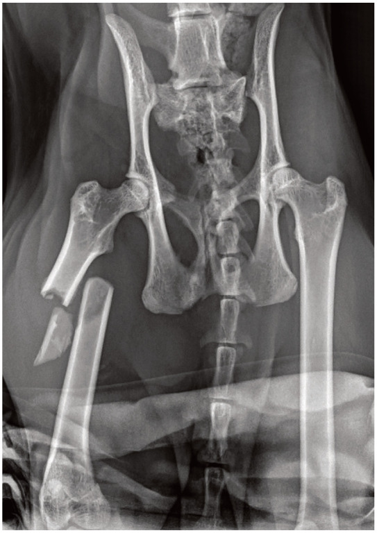
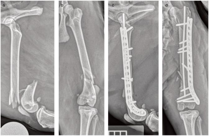
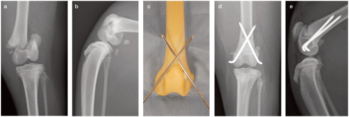
Practical relevance: The femur is the most commonly fractured bone in cats. Femoral fractures usually result from high-velocity trauma such as a road traffic accident or fall from a height and, as such, are associated with a wide variety of concurrent injuries. The initial focus of treatment should always be on assessment and stabilisation of the major body systems. Once any concurrent injuries have been addressed, all femoral fractures need surgical stabilisation, with the notable exception of greenstick fractures in very young cats, which can heal with cage rest alone. A number of different surgical options are available depending on the fracture type, location, equipment, surgeon experience and owner finances.

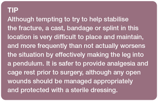
Clinical challenges: Femoral fractures can vary hugely in complexity and the small size of feline bones can limit the choice of implants. Furthermore, cats can present unique challenges in the postoperative period due to their active nature and the limited means to control their exercise level.

Audience: This review is aimed at general and feline-specific practitioners who have some experience of feline orthopaedics, as well as those simply wishing to expand their knowledge.

Aims: The aim of this review is to help clinicians assess, plan and manage feline femoral fractures. It provides an overview of diagnostic imaging and a discussion of a range of suitable surgical options, including the principles of different types of fixation. It also highlights cat-specific issues, approaches and implants pertinent to the management of these cases.

Evidence base: A number of original articles and textbook chapters covering many aspects of femoral fractures in cats and dogs have been published. Where possible, this review draws on information from key feline research and, where necessary, extrapolates from relevant canine literature. The authors also offer practical guidance based on their own clinical experience.

Keywords: Femur; capital femoral physis; distal femoral physis; femoral; fracture; trauma.


Conflict of interest statement

The authors declared no potential conflicts of interest with respect to the research, authorship, and / or publication of this article.

Figures

Figure 1
Figure 1
(a) Craniocaudal and (b) mediolateral radiographic views of a feline femur with the major landmarks indicated. Note the mineralisation of the medial fabella, which many cats will not have. The lateral fabella, which is always mineralised in cats, is indicated by the dotted pink circle in (a). (c) Magnified view of a section of the femoral diaphysis demonstrating how to measure the femoral diameter and medullary canal diameter. Note that it is important always to include a marker for referencing accurate size measurements; a ruler can be seen in the bottom left corner of image (b), but the remainder of the markers have been lost from these radiographs during image cropping
Figure 2
Figure 2
(a) Ventrodorsal frog-leg radiographic view of a feline pelvis showing avulsion of the ischial tuberosity (short arrow) but no other clear injuries. (b) Ventrodorsal extended-leg radiographic view of the same feline pelvis showing again the avulsion of the ischial tuberosity (short arrow) but most importantly also a proximal femoral physeal fracture (long arrow), which was not evident on the ventrodorsal frog-leg radiograph
Figure 3
Figure 3
Musculature of the proximal hindlimb of the cat. Note that the gluteal muscles may be relatively larger in cats than dogs and the caudofemoralis muscle, not present in the dog, is interposed between the superficial gluteal and biceps femoris muscles
Figure 4
Figure 4
(a) Model showing the ideal overlap position of two Kirschner wires for femoral neck or proximal femoral physeal fractures. Ideally the pins should be parallel to the femoral neck and start distal to the third trochanter. (b) Lateral and (c) ventrodorsal extended and (d) frog-leg views showing the repair of a Salter-Harris type I femoral head fracture with two parallel Kirschner wires
Figure 5
Figure 5
(a) Ventrodorsal radiographic view of a kitten with polytrauma including a left hip luxation, left sacroiliac luxation, right ischial fractures and left ischial tuberosity avulsion. The greater trochanter of the left femur does not have a normal appearance (arrow); however, this could be due to rotation. At surgery a trochanteric avulsion was also found. (b) Ventrodorsal radiographic view of the pelvis of the same kitten following repair of the greater trochanteric fracture with two Kirschner wires and a polydioxanone tension band. Note that the left sacroiliac luxation has also been stabilised via placement of a sacroiliac screw
Figure 6
Figure 6
Ventrodorsal radiographic view of a feline femur showing an intramedullary pin that was placed as a sole means of fixation for a simple mid-diaphyseal fracture. The fracture subsequently displaced and then fissures developed in line with the axis of the pin
Figure 7
Figure 7
Femoral model showing (a) a proximal bird’s-eye view of the femur with the location (circle) for initiating intramedullary pin placement into the trochanteric fossa, and (b) lateral and (c) cranial views of the femur demonstrating alignment of an intramedullary pin
Figure 8
Figure 8
Three-dimensional CT reconstruction of a feline femur shown from (a) craniocaudal, (b) lateromedial, (c) caudocranial and (d) mediolateral directions. Slight anteversion (pointing forwards) of the femoral head can be seen, which is typical in cats. The linea aspera, identified by the arrow on images (b) and (c), can be a helpful visual aid when assessing for accurate fracture reduction and/or anatomical alignment. The long and relatively straight femoral canal with minimal metaphyseal flare is typical in cats. Also note this cat has both medial and lateral fabellae
Figure 9
Figure 9
Intraoperative use of a fracture distractor as an aid to realign a femoral fracture in a cat. The fracture distractor is placed with one full pin in the distal femoral condyle and one in the greater trochanter. The distractor is then used to slowly drive the two ends of the fracture apart while maintaining alignment
Figure 10
Figure 10
Plate options for a feline femur laid out to demonstrate the different screw hole densities. From left to right the plates are: a 2.0 mm dynamic compression plate, a 2.4 mm locking compression plate (LCP), a 2.7 mm LCP and a 2.0 mm/2.7 mm veterinary cuttable plate
Figure 11
Figure 11
Plate options for a feline femur as demonstrated on a model femur: (a) 2.0 mm/2.7 mm veterinary cuttable plate, (b) 2.4 mm locking compression plate (LCP), (c) 2.7 mm LCP and (d) a plate-rod configuration
Figure 12
Figure 12
Models showing the three options for plate application and the subsequent effect on the implant’s function and the forces acting through the bone and implant. (a) Compression (fracture fragments are compressed and the fracture has load sharing). (b) Neutralisation (lag screw placed across the fracture; the fracture ends are compressed by the lag screw but not by the plate and, as such, the fracture still has some load sharing). (c) Bridging (the fracture ends are not compressed and there is no load sharing; the plate takes all of the load)
Figure 13
Figure 13
Fixator pin options include those that are (a) end-threaded positive profile, (b) end-threaded negative profile and (c) mid-threaded positive profile. Single clamp options include (d) Kirschner-Ehmer (KE)-style clamps and (e) IMEX-SK-style clamps. Double clamp options include (f) IMEX-SK-style clamps and (g) KE-style clamps. (h) Lateral radiographic view of a feline femur showing measurement of the isthmus (narrowest part) for estimation of appropriate fixator pin sizes (femoral diameter) and intramedullary pin sizes (medullary canal diameter)
Figure 14
Figure 14
(a) Lateral femoral radiograph showing a long spiral fracture of the mid-femoral diaphysis in a cat. (b) Craniocaudal femoral radiograph of the cat in (a) following stabilisation with an intramedullary pin tied into a type Ia external skeletal fixator using two end-threaded positive profile pins proximally and distally. (c) Lateral femoral radiograph of a different cat showing a grade II open distal diaphyseal femoral fracture (note the gas in the soft tissues). (d) Lateral femoral radiograph of the cat in (c) following stabilisation with an epoxy putty free-form fixator, combining a tied-in intramedullary pin
Figure 15
Figure 15
Ventrodorsal radiographic view of a feline pelvis showing a mid- to proximal third diaphyseal femoral segmental fracture
Figure 16
Figure 16
(a) Ventrodorsal radiographic view of a feline pelvis showing a short oblique slightly comminuted mid-diaphyseal femoral fracture. A historical femoral neck fracture, stabilised with three pins, and repaired with two pins and a figure-of-eight tension band wire) is also evident. (b) Craniocaudal radiographic view of the femur in (a) following surgical stabilisation. A pin was placed across the fracture, and a 2.4 mm locking compression plate was applied to the lateral aspect with a mixture of cortical and locking screws
Figure 17
Figure 17
(a) Lateral and (b) craniocaudal radiographic views of a highly comminuted and fissured diaphyseal femoral fracture. Postoperative (c) lateral and (d) craniocaudal radiographic views of the cat in (a,b) showing a plate-rod repair, whereby a 1.6 mm Kirschner wire has been placed as an intramedullary pin and a lateral 2.4 mm locking compression plate has been contoured and applied. Note that no attempt has been made to reconstruct the fracture
Figure 18
Figure 18
(a) Lateral and (b) craniocaudal radiographic views of a comminuted and fissured supracondylar femoral fracture. (c) Lateral and (d) craniocaudal radiographic views of the cat in (a,b) following stabilisation with an intramedullary pin and a J-shaped hockey-stick plate (plate-rod construct), which allows for more screws to be placed in the short and backward-curving distal fragment. The plate has been cut to length proximally using large pin cutters
Figure 19
Figure 19
Preoperative (a) caudocranial and (b) mediolateral radiographic views of a feline stifle, showing a Salter-Harris type I distal femoral physeal fracture. (c) Model showing correct cross-pin placement and alignment. Postoperative (d) caudocranial and (e) mediolateral radiographic views of the stifle in (a,b) showing fracture reduction and crossed Kirschner wire pin placement (1.2 mm pins), with the ends bent over. Note the pins must cross above the fracture and the trochar tip must exit through the femoral cortex
Figure 20
Figure 20
Craniocaudal radiographic projection of a cat with a Salter-Harris type IV femoral condylar fracture
None
None
None
None
None
None
None
None
None
None
None
None
None
None

Similar articles

Cited by

References

    1. Hill FW. A survey of bone fractures in the cat. J Small Anim Pract 1977; 18: 457-463. - PubMed
    1. Bookbinder PF, Flanders JA. Characteristics of pelvic fracture in the cat. Vet Comp Orthop Traumatol 1992; 37: 122-127.
    1. Voss K, Langley-Hobbs SJ, Montavon PM. Femur. In: Montavon PM, Voss K, Langley-Hobbs SJ. (eds). Feline orthopaedic surgery and musculoskeletal disease. Edinburgh: Saunders Elsevier, 2009, pp 455-474.
    1. Meeson R, Corr S. Management of pelvic trauma: neurological damage, urinary tract disruption and pelvic fractures. J Feline Med Surg 2011; 13: 347-361. - PMC - PubMed
    1. Meeson RL, Geddes AT. Management and long-term outcome of pelvic fractures: a retrospective study of 43 cats. J Feline Med Surg 2017; 19: 36-41. - PMC - PubMed