. 2022 Jun 20;61(25):e202204052.
doi: 10.1002/anie.202204052. Epub 2022 Apr 25.

Rational Design of a Novel Tubulin Inhibitor with a Unique Mechanism of Action

Affiliations

Rational Design of a Novel Tubulin Inhibitor with a Unique Mechanism of Action

Tobias Mühlethaler et al. Angew Chem Int Ed Engl. .

Abstract

In this study, we capitalized on our previously performed crystallographic fragment screen and developed the antitubulin small molecule Todalam with only two rounds of straightforward chemical synthesis. Todalam binds to a novel tubulin site, disrupts microtubule networks in cells, arrests cells in G2/M, induces cell death, and synergizes with vinblastine. The compound destabilizes microtubules by acting as a molecular plug that sterically inhibits the curved-to-straight conformational switch in the α-tubulin subunit, and by sequestering tubulin dimers into assembly incompetent oligomers. Our results describe for the first time the generation of a fully rationally designed small molecule tubulin inhibitor from a fragment, which displays a unique molecular mechanism of action. They thus demonstrate the usefulness of tubulin-binding fragments as valuable starting points for innovative antitubulin drug and chemical probe discovery campaigns.

Keywords: Fragments; Microtubule-Targeting Agents; Molecular Mechanism of Action; Rational Drug Design; Tubulin.


Conflict of interest statement

The authors declare no conflict of interest.

Figures

Figure 1
Figure 1
Fragment selection. A) Location of the site sID βαII (volume representation) at the βTub1‐αTub2 interdimer interface of the T2R‐TTL complex. For simplicity, the RB3 and TTL chains are not shown. The two α‐ and two β‐tubulin monomers in the T2R‐TTL complex are shown in grey and white ribbon representation, respectively. B) Superimposition of the F04, F36, and F41 binding poses (PDB IDs 5S4O, 5S5K, and 5S5P, respectively). Secondary structure elements of tubulin are labeled in blue. Carbon atoms are colored in green for F04, orange for fragment F36, and light blue for fragment F41. Nitrogen atoms are colored in blue, oxygen atoms in red, sulfur atoms in yellow, and fluorine atoms in light green. C) Chemical structures of fragments F04, F36, and F41. D) Schematic representation of F04 bound to site sID βαII. Residues forming the binding site are shown in green for hydrophobic, in light blue for polar, and in dark blue for charged residues. The interacting residues αL242 and αL252 are not shown because they are located below the ligand. Hydrogen bonds are indicated with dashed and π‐stacking interactions with solid black lines. Red dots represent crystallographic water molecules.
Scheme 1
Scheme 1
Reaction conditions for Hantzsch synthesis (35, 810 and 1422), reductive amination (1, 2), and amide derivatives (1113). Reagents and conditions: (i) 4‐acetamidophenacyl bromide (1  equiv), thiourea (1.1  equiv), EtOH, 1 h, reflux, yield 27–96 %; (ii) N‐(4‐(2‐aminothiazol‐4‐yl)phenyl)acetamide (1  equiv), aldehyde (2 equiv), AcOH (5 equiv), NaBH(OAc)3 (2.8 equiv), DCE, 48 h, rt, yield 24–56 % (iii) 2‐bromo‐1‐(4‐nitrophenyl)ethanone (1 equiv) and N‐(3‐fluorophenyl)thiourea (1.1 equiv), EtOH, 1 h, reflux, yield 72 %; (iv) Fe (15 equiv), 24.5 % HCl (2 equiv), iPrOH, 1 h, 60 °C, yield 36 %; (v) acyl chloride (1.1 equiv), DIPEA (2 equiv), DCM, 30 min, 0 °C, 1–2 h, rt, yield 52–71 %.
Scheme 2
Scheme 2
Reaction conditions for the synthesis of compounds 6 and 7. Reagents and conditions: (i) N‐(4‐(2‐aminothiazol‐4‐yl)phenyl)acetamide (1 equiv), benzoylchloride (2 equiv), triethylamine (2 equiv), anhydrous DCM, 0 °C to rt, N2, 16 hours, yield 25 %; (ii) N‐(4‐(2‐aminothiazol‐4‐yl)phenyl)acetamide (1 equiv), 2‐methanesulfonyl chloride (1 equiv), anhydrous DCM, pyridine, 0 °C to rt, N2, 6 hours, yield 70 %.
Figure 2
Figure 2
Structural basis of 4 and its meta‐, ortho‐, and para‐fluorinated derivatives. A) Superimposition of the binding site of the tubulin‐4 complex structure onto the ones of tubulin‐F04 (green) and tubulin‐F41 (blue). B) Superimposition of the binding site of the tubulin‐10 complex structure onto the ones of tubulin‐F04 (green) and tubulin‐F41 (blue). C) Superimposition of the binding site of the tubulin‐9 complex structure onto the ones of tubulin‐F04 (green) and tubulin‐F36 (orange). D) Superimposition of the binding site of the tubulin‐8 complex structure onto the ones of tubulin‐F04 (green) and tubulin‐F41 (blue). The α‐ and β‐tubulin monomers are shown in gray and white ribbon representation, respectively. Residues forming the binding site are shown in stick representation and are labeled in black. The same atom color scheme as in Figure 1B has been used. Hydrogen bonds are shown as dashed black lines.
Figure 3
Figure 3
Structural basis of Todalam. A) Binding mode of Todalam as seen in the crystal structure of the tubulin‐Todalam complex superimposed onto the one of the tubulin‐9 complex. The α‐ and β‐tubulin monomers are shown in gray and white ribbon representation, respectively. Residues forming the binding site are shown in stick representation and are labeled in black. The same atom color scheme as in Figure 1B has been used. Hydrogen bonds are shown as dashed black lines. B) The Todalam site is shown in surface representation and colored with a gradient from red for a hydrophobic to white for a hydrophilic environment. The chemical structure of Todalam is shown on the bottom right of the panel.
Figure 4
Figure 4
Effect of Todalam on tubulin and microtubules in vitro. A) Inhibition of tubulin polymerization assessed by microtubule pelleting assays performed in the presence of the indicated increasing amounts of Todalam or control compounds at 10 μM. Tubulin concentration: 30 μM. For raw data, see Figure S7A. B) Negative staining electron micrographs of tubulin incubated together with 150 μM Todalam (gallery) or without the compound (overview micrograph). Tubulin concentration: 20 μM. Scale bars, 50 nm.
Figure 5
Figure 5
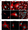
Effect of Todalam on microtubules in cells. A) Representative fluorescence confocal microscopy images of HMF3a, MDAMB 231, and HeLa cells that were treated either with 0.5 % DMSO (control, upper panels) or 20 μM Todalam (lower panels) for 4 hours. Microtubules are immunostained with an anti α‐tubulin antibody and are colored in red; the cell nuclei are stained with DAPI (4′,6‐diamidin‐2‐phenylindole) and are colored in blue. B) Drug wash out experiments. Representative confocal images of HMF3a cells that were treated with 20 μM Todalam for 4 hours, subsequently washed with culture media, and left to recover for 16 hours. The immunostaining assay is identical to the one shown in panel A. Scale bars, 20 μm.
Figure 6
Figure 6
Molecular mechanism of action of Todalam. A, B) Superimposition of the α‐tubulin (A) and β‐tubulin (B) half sites of the Todalam pocket in the curved and straight conformation of the tubulin dimer. The curved conformational states of the α‐ and β‐tubulin subunits are shown in grey and white ribbon representation, respectively. The straight states of tubulin monomer structures were taken from PDB ID 6DPV and are colored according to their three domains: N‐terminal domain in yellow (residues 1–205), intermediate domain in orange (residues 206–384), and C‐terminal domain in red (residues 385–440). For the superposition of the curved and straight conformational states of α‐ and β‐tubulin, the central strands αS1, αS2, αS4, αS5, and αS6, or βS1, βS2, βS4, βS5, and βS6 were used, respectively. Secondary structure elements are labelled in blue. For simplicity, strand αS1 that is not involved in the molecular mechanism of action of Todalam was removed in panel A. The black arrow in panel A indicates the movement of helix αH8 during the curved‐to‐straight conformational transition of the tubulin dimer. C) Schematic illustration of the molecular mechanism of action of Todalam on tubulin and microtubules. Upper panel: Tubulin assembly in the absence of Todalam, highlighting the shift of the αH8 helix of α‐tubulin required for proper accommodation of the incoming tubulin dimer. Lower Panel: The presence of Todalam blocks the shift of the αH8 helix, thereby preventing microtubule growth and promoting the formation of ring‐like oligomers.

References

    1. None
    1. Yang C. H., Horwitz S. B., Int. J. Mol. Sci. 2017, 18, 1733;
    1. Miltenburg N. C., Boogerd W., Cancer Treat. Rev. 2014, 40, 872–882; - PubMed
    1. Kavallaris M., Nat. Rev. Cancer 2010, 10, 194–204. - PubMed
    1. None

Publication types

LinkOut - more resources