. 2022 Apr 12;145(15):1123-1139.
doi: 10.1161/CIRCULATIONAHA.121.056817. Epub 2022 Apr 11.

Prevalence, Characteristics, and Outcomes of COVID-19-Associated Acute Myocarditis

Affiliations

Prevalence, Characteristics, and Outcomes of COVID-19-Associated Acute Myocarditis

Enrico Ammirati et al. Circulation. .

Abstract

Background: Acute myocarditis (AM) is thought to be a rare cardiovascular complication of COVID-19, although minimal data are available beyond case reports. We aim to report the prevalence, baseline characteristics, in-hospital management, and outcomes for patients with COVID-19-associated AM on the basis of a retrospective cohort from 23 hospitals in the United States and Europe.

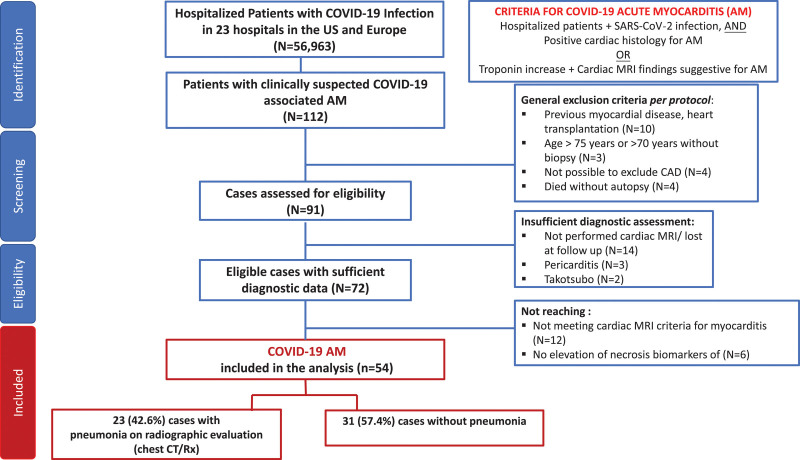
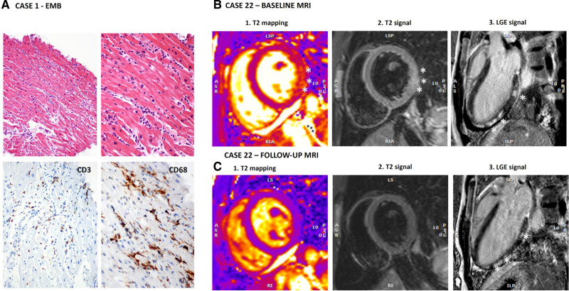
Methods: A total of 112 patients with suspected AM from 56 963 hospitalized patients with COVID-19 were evaluated between February 1, 2020, and April 30, 2021. Inclusion criteria were hospitalization for COVID-19 and a diagnosis of AM on the basis of endomyocardial biopsy or increased troponin level plus typical signs of AM on cardiac magnetic resonance imaging. We identified 97 patients with possible AM, and among them, 54 patients with definite/probable AM supported by endomyocardial biopsy in 17 (31.5%) patients or magnetic resonance imaging in 50 (92.6%). We analyzed patient characteristics, treatments, and outcomes among all COVID-19-associated AM.

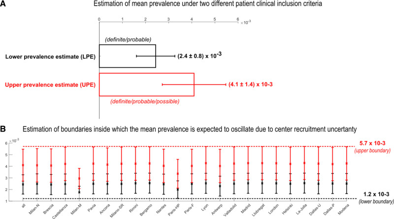
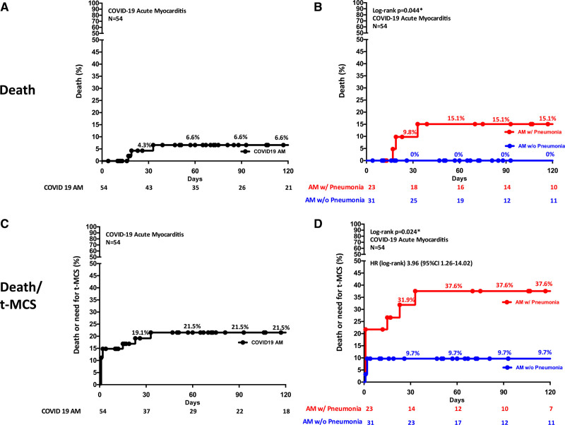
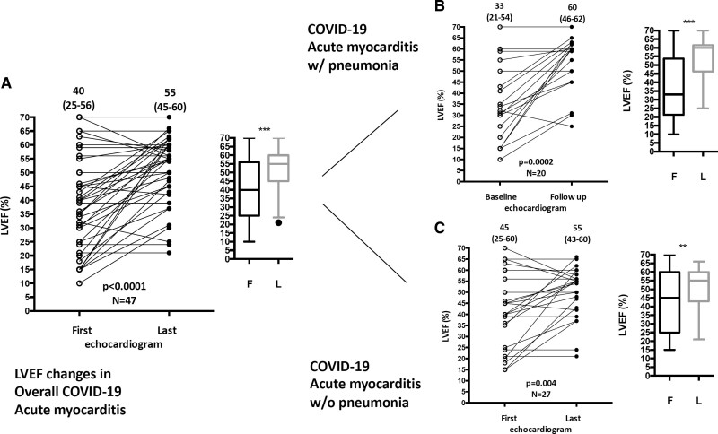
Results: AM prevalence among hospitalized patients with COVID-19 was 2.4 per 1000 hospitalizations considering definite/probable and 4.1 per 1000 considering also possible AM. The median age of definite/probable cases was 38 years, and 38.9% were female. On admission, chest pain and dyspnea were the most frequent symptoms (55.5% and 53.7%, respectively). Thirty-one cases (57.4%) occurred in the absence of COVID-19-associated pneumonia. Twenty-one (38.9%) had a fulminant presentation requiring inotropic support or temporary mechanical circulatory support. The composite of in-hospital mortality or temporary mechanical circulatory support occurred in 20.4%. At 120 days, estimated mortality was 6.6%, 15.1% in patients with associated pneumonia versus 0% in patients without pneumonia (P=0.044). During hospitalization, left ventricular ejection fraction, assessed by echocardiography, improved from a median of 40% on admission to 55% at discharge (n=47; P<0.0001) similarly in patients with or without pneumonia. Corticosteroids were frequently administered (55.5%).

Conclusions: AM occurrence is estimated between 2.4 and 4.1 out of 1000 patients hospitalized for COVID-19. The majority of AM occurs in the absence of pneumonia and is often complicated by hemodynamic instability. AM is a rare complication in patients hospitalized for COVID-19, with an outcome that differs on the basis of the presence of concomitant pneumonia.

Keywords: COVID-2019; MRI; SARS-CoV-2; cardiac; myocarditis; outcome.


Figures

Figure 1.
Figure 1.
Flow diagram illustrating screening and inclusion criteria. AM indicates acute myocarditis; CAD, coronary artery disease; CT, computed tomography; MRI, magnetic resonance imaging; and Rx‚ radiograph.
Figure 2.
Figure 2.
Estimation of lower prevalence estimate (LPE) and upper prevalence estimate (UPE) of acute myocarditis among hospitalized patients with COVID-19. A, Mean LPE and mean UPE computed with the respective CIs on the 23 centers. B, Mean LPE and mean UPE with the respective CIs iteratively computed on 22 centers (by means of leave-1-out procedure). The red dashed line at the top (which is the maximum level reached by the CIs) and the black dashed line at the bottom (which is the minimum level reached by the CIs) represent the boundaries inside which the estimation of the sample mean prevalence is expected to occur. Dallas-P indicates Dallas-Parkland health & Hospital System; Dallas-U, Dallas-University of Texas Southwestern Medical Center; Milan-M, Milan Monzino; Milan-N, Milan Niguarda hospital; Milano-SR, Milano San Raffaele hospital; Paris-F, Paris-Foch; and Paris-HP, Paris-Hôpital Pitié–Salpêtrière.
Figure 3.
Figure 3.
Histological findings and cardiac magnetic resonance imaging (MRI) of 2 patients with COVID-19–associated acute myocarditis. A, Endomyocardial biopsy (EMB) findings from a 20-year-old male patient (case 1 in Table S2) show inflammatory infiltrates in the myocardium (upper images, hematoxylin and eosin images at 100× and 200× magnification) and positive CD3 and CD68 immunohistochemical stains (lower images, images at 200× magnification) revealing CD3+T-lymphocytes ≥7 cells/mm2 and CD68+macrophages ≥4 cells/mm2 consistent with myocarditis on the basis of European Society of Cardiology criteria. B, Baseline and follow-up cardiac MRI images at 1.5 Tesla of a 16-year-old boy (case 22) admitted to the hospital with acute myocarditis without pneumonia. Cardiac MRI at baseline fulfilled the 2018 Lake Louise Criteria for myocarditis because signs of both myocardial edema and nonischemic myocardial injury were present. T2-weighted images showed patchy areas of increased T2 signal intensity. In B, both T2 mapping (1) and short-tau inversion recovery (STIR) T2-weighted imaging (2) showed an area of increased signal intensity (SI) in the basal inferolateral wall (asterisks); in this area, T2 mapping value was 58 ms, whereas it was 43 ms in the septum, and the ratio between myocardial and skeletal muscle SI was elevated at 2.5. Postcontrast images (3) showed patchy late gadolinium enhancement (LGE) with nonischemic pattern in the inferolateral wall (asterisks). Pericardial effusion was also evident at cine images, whereas global systolic function was preserved (left ventricular ejection fraction [LVEF] 63%, Video S1). In C, follow-up images of the same patient at 6 months are shown. Compared with the scan acquired in the acute phase, there were no signs of myocardial edema: in the basal inferolateral wall, T2 mapping value decreased to 44 ms (1), and SI ratio between the myocardium and skeletal muscle at STIR T2-weighted images was <2 (2). Patchy LGE (asterisks) of the inferolateral wall persisted, although reduced (3). The pericardial effusion was still present, and systolic ventricular function improved (LVEF 69%; see Video S2). Of note, indexed myocardial mass also reduced from baseline to follow-up from 85 to 78 g/m2. Increased mass in the acute phase can represent an indirect sign of myocardial edema.
Figure 4.
Figure 4.
Kaplan -Meier estimates of 120-day overall mortality and mortality or need for temporary mechanical circulatory support (t-MCS) in patients with COVID-19–associated acute myocarditis (AM). A, Mortality in the whole study population with COVID-19 am and (B) Mortality in patients with COVID-19 am with (w/) pneumonia on the basis of radiographic examinations compared with patients with COVID-19 am without (w/o) pneumonia. Three noncardiac deaths occurred: 2 deaths caused by septic shock and 1 caused by hemorrhagic stroke. C, Mortality or need for t-MCS in the whole study population with COVID-19 am and (D) Mortality or need for t-MCS in patients with COVID-19 am with pneumonia compared with patients with COVID-19 am without pneumonia. Among 10 patients who received a t-MCS, 2 died on support (1 septic shock and 1 hemorrhagic stroke). HR indicates hazard ratio. *Indicates statistically significant with P<0.05.
Figure 5.
Figure 5.
Changes in left ventricular ejection fraction (LVEF) during hospitalization in patients with COVID-19 acute myocarditis. A, Echocardiographic data of LVEF at admission and discharge in the entire population of COVID-19 acute myocarditis (available data, n=47 of 54). B, COVID-19 acute myocarditis with (w/) pneumonia (available data, n=20 of 23), and (C) COVID-19 acute myocarditis without (w/o) pneumonia (available data, n=27 of 31). Wilcoxon matched-pair signed-rank test was used for comparisons. The dot plots indicate the median and first and third quartile LVEF at baseline and at follow-up in each group. F indicates first; and L, last. **Indicates P<0.01; ***, P<0.001.

References

    1. Berlin DA, Gulick RM, Martinez FJ. Severe Covid-19. N Engl J Med. 2020;383:2451–2460. doi: 10.1056/NEJMcp2009575 - PubMed
    1. Guo J, Huang Z, Lin L, Lv J. Coronavirus disease 2019 (COVID-19) and cardiovascular disease: a viewpoint on the potential influence of angiotensin-converting enzyme inhibitors/angiotensin receptor blockers on onset and severity of severe acute respiratory syndrome coronavirus 2 infection. J Am Heart Assoc. 2020;9:e016219. doi: 10.1161/JAHA.120.016219 - PMC - PubMed
    1. Lala A, Johnson KW, Januzzi JL, Russak AJ, Paranjpe I, Richter F, Zhao S, Somani S, Van Vleck T, Vaid A, et al. ; Mount Sinai COVID Informatics Center. Prevalence and impact of myocardial injury in patients hospitalized with COVID-19 infection. J Am Coll Cardiol. 2020;76:533–546. doi: 10.1016/j.jacc.2020.06.007 - PMC - PubMed
    1. Li C, Jiang J, Wang F, Zhou N, Veronese G, Moslehi JJ, Ammirati E, Wang DW. Longitudinal correlation of biomarkers of cardiac injury, inflammation, and coagulation to outcome in hospitalized COVID-19 patients. J Mol Cell Cardiol. 2020;147:74–87. doi: 10.1016/j.yjmcc.2020.08.008 - PMC - PubMed
    1. Alvarez-Garcia J, Jaladanki S, Rivas-Lasarte M, Cagliostro M, Gupta A, Joshi A, Ting P, Mitter SS, Bagiella E, Mancini D, et al. . New heart failure diagnoses among patients hospitalized for COVID-19. J Am Coll Cardiol. 2021;77:2260–2262. doi: 10.1016/j.jacc.2021.03.006 - PMC - PubMed

Publication types