

Thrombus characteristics evaluated by acute optical coherence tomography in ST elevation myocardial Infarction
- PMID: 35404941
- PMCID: PMC9000063
- DOI: 10.1371/journal.pone.0266634
Thrombus characteristics evaluated by acute optical coherence tomography in ST elevation myocardial Infarction
Abstract
Aims: ST elevation myocardial infarction (STEMI) is caused by an occlusive thrombosis of a coronary artery. We wanted to assess if the thrombus can be characterized according to erythrocyte content and age using intravascular optical coherence tomography (OCT) in a clinical setting.
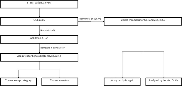
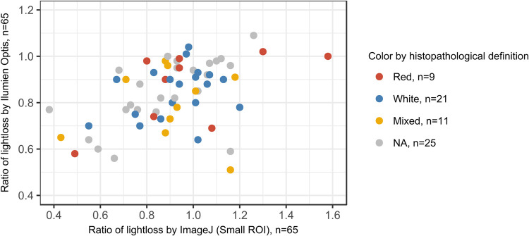
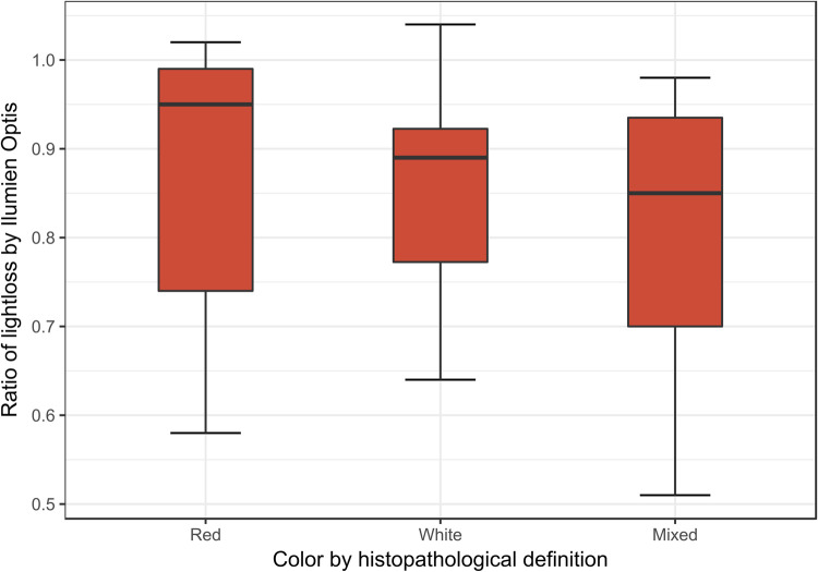
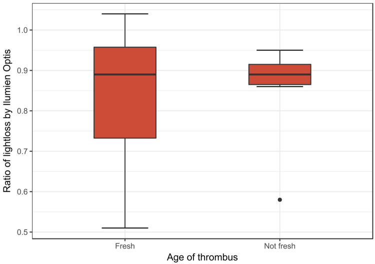
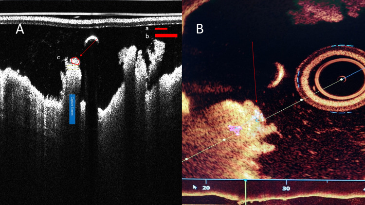
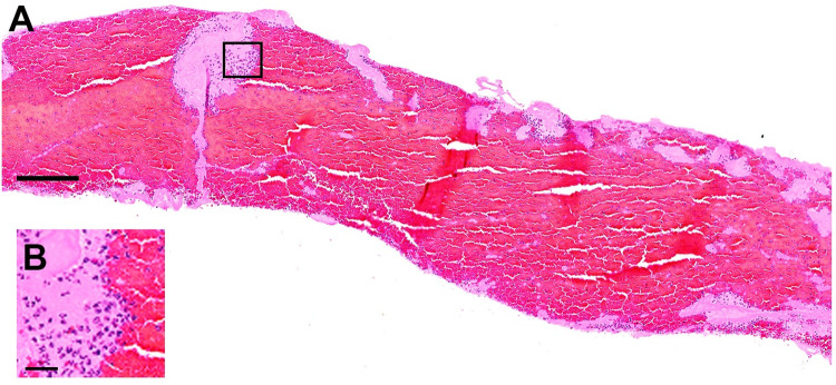
Methods and results: We performed manual thrombus aspiration in 66 STEMI patients. OCT was done of the thrombus remnants after aspiration. A light intensity ratio was measured through the thrombus. Forty two of the aspirates had thrombus which could be analyzed histomorphologically for analysis of erythrocyte and platelet content, and to determine the age of thrombus as fresh, lytic or organized. There were 11 red, 21 white and 10 mixed thrombi. Furthermore, 36 aspirates had elements of fresh, 7 of lytic and 8 of organized thrombi. There was no correlation between colour and age. OCT appearance could not predict erythrocyte or platelet content. The light intensity ratios were not significantly different in fresh, lytic or organized thrombi.
Conclusion: OCT could not differentiate between red and white thrombi, nor determine thrombus age.
Conflict of interest statement
The authors have declared that no competing interests exist.
Figures
References
-
- Ibanez B, James S, Agewall S, Antunes MJ, Bucciarelli-Ducci C, Bueno H, et al. 2017 ESC Guidelines for the management of acute myocardial infarction in patients presenting with ST-segment elevation: The Task Force for the management of acute myocardial infarction in patients presenting with ST-segment elevation of the European Society of Cardiology (ESC). European heart journal. 2018;39(2):119–77. doi: 10.1093/eurheartj/ehx393 - DOI - PubMed
-
- George JC, Dangas GD, American Heart A, American College of C. 2009 Focused updates to guidelines in ST-elevation myocardial infarction and percutaneous coronary intervention: application to interventional cardiology. JACC Cardiovascular interventions. 2010;3(2):256–8. doi: 10.1016/j.jcin.2010.01.004 - DOI - PubMed
-
- Rittersma SZ, van der Wal AC, Koch KT, Piek JJ, Henriques JP, Mulder KJ, et al. Plaque instability frequently occurs days or weeks before occlusive coronary thrombosis:a pathological thrombectomy study in primary percutaneous coronary intervention. Circulation.2005;111(9):1160–5. doi: 10.1161/01.CIR.0000157141.00778.AC - DOI - PubMed
-
- Kramer MC, van der Wal AC, Koch KT, Ploegmakers JP, van der Schaaf RJ, Henriques JP, et al. Presence of older thrombus is an independent predictor of long-term mortality in patients with ST-elevation myocardial infarction treated with thrombus aspiration during primary percutaneous coronary intervention. Circulation. 2008;118(18):1810–6. doi: 10.1161/CIRCULATIONAHA.108.780734 - DOI - PubMed
MeSH terms
LinkOut - more resources
Full Text Sources
Medical
