. 2022 Aug 25;31(17):2934-2950.
doi: 10.1093/hmg/ddac085.

The microRNA processor DROSHA is a candidate gene for a severe progressive neurological disorder

Collaborators, Affiliations

The microRNA processor DROSHA is a candidate gene for a severe progressive neurological disorder

Scott Barish et al. Hum Mol Genet. .

Abstract

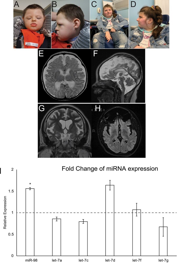
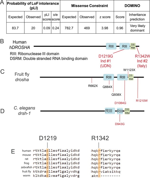
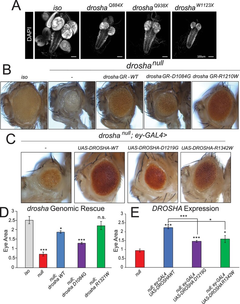
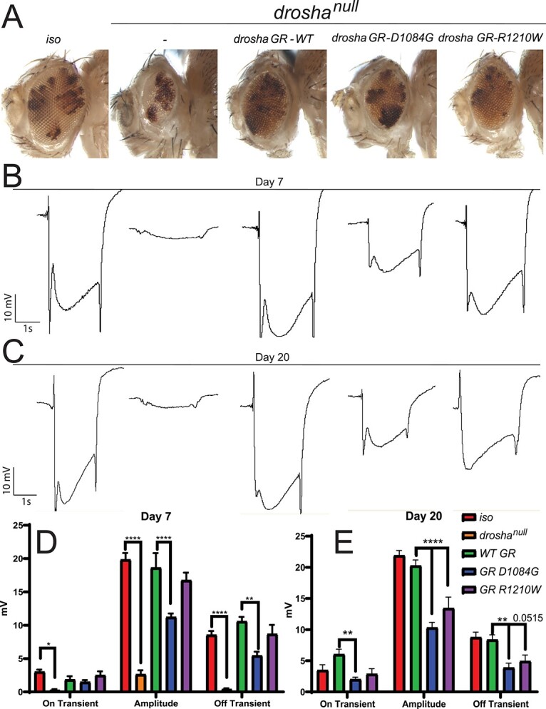
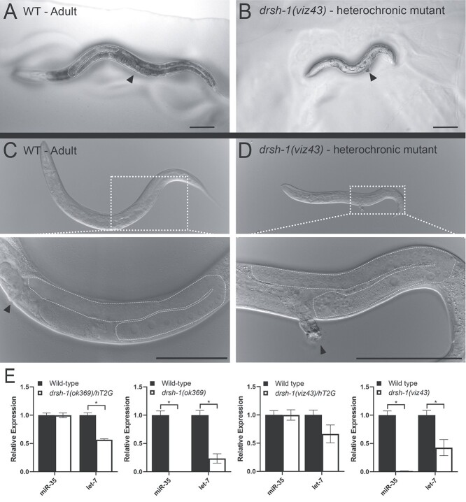
DROSHA encodes a ribonuclease that is a subunit of the Microprocessor complex and is involved in the first step of microRNA (miRNA) biogenesis. To date, DROSHA has not yet been associated with a Mendelian disease. Here, we describe two individuals with profound intellectual disability, epilepsy, white matter atrophy, microcephaly and dysmorphic features, who carry damaging de novo heterozygous variants in DROSHA. DROSHA is constrained for missense variants and moderately intolerant to loss-of-function (o/e = 0.24). The loss of the fruit fly ortholog drosha causes developmental arrest and death in third instar larvae, a severe reduction in brain size and loss of imaginal discs in the larva. Loss of drosha in eye clones causes small and rough eyes in adult flies. One of the identified DROSHA variants (p.Asp1219Gly) behaves as a strong loss-of-function allele in flies, while another variant (p.Arg1342Trp) is less damaging in our assays. In worms, a knock-in that mimics the p.Asp1219Gly variant at a worm equivalent residue causes loss of miRNA expression and heterochronicity, a phenotype characteristic of the loss of miRNA. Together, our data show that the DROSHA variants found in the individuals presented here are damaging based on functional studies in model organisms and likely underlie the severe phenotype involving the nervous system.


Figures

Figure 1
Figure 1
Two individuals with DROSHA variants show facial dysmorphia, microcephaly and white matter atrophy. (A and B) Individual 1 at 3 years, 11 months. (C and D) Individual 2 at 23 years. (E and F) Axial (E) and sagittal (F) T2 weighted MR images from Individual 1 obtained at age 5 weeks show global atrophy. (G and H) Coronal T2 (G) and axial FLAIR (H) MR images from patient 2 obtained at age 17 years show global atrophy. (I) Expression of a panel of miRNAs in DROSHAD1219G fibroblasts. miRNA expression was assayed using TaqMan assays and compared to control fibroblasts. miR98 expression was significantly upregulated in DROSHAD1219G fibroblasts. *P < 0.5.
Figure 2
Figure 2
DROSHA is variant constrained and its protein structure is highly conserved. (A) Bioinformatics analysis of DROSHA genetic variants from the gnomAD (59). DROSHA has an observed/expected (o/e) score of 0.24, which suggests that is not tolerant to loss-of-function variants. It also has a high missense constraint score of 3.98 and is predicted to be very likely dominant by DOMINO (106). (B–D) Protein structure of human (B), fruit fly (C) and C. elegans (D). DROSHA proteins are highly conserved in the three species, with all proteins containing two Ribonuclease III domains (RIII) and a single DSRM domain. The residues affected by the patient variants are shown in red in (B) and their corresponding residues are shown in (C–E). Drosha truncation mutations that have been annotated as null alleles are labeled in black in (C). (E) Conservation of affected patient residues. Both Individual 1 (left) and 2’s (right) variants affect residues that are conserved between humans and flies but only Individual 1’s residue is conserved in all three species.
Figure 3
Figure 3
DROSHA variants damage protein function and are partially able to rescue fly eye/head size defects. (A) Third instar larval brains stained with DAPI to show nuclei. Control fly larvae (first panel) display a wild-type brain and attached imaginal discs (first panel). Drosha null allele mutants show dramatically reduced brains as well as a loss of the attached imaginal discs (second–fourth panels). The VNC that can be seen posterior to the two brain lobes is unaffected. (B) Eye-specific droshaW1123X clones were generated using the ey-GAL4 UAS-FLP/FRT system (74). A fly with clones of the isogenized FRT42D chromosome (control, first panel) displays wild-type eye size. droshaW1123X (referred to as droshanull in the images for simplicity, second panel) clones cause both the eye and head size to be reduced. Introduction of a wild-type GR (drosha GR-WT, third panel) construct is able to partially rescue eye and head size defect. Introduction of a GR construct carrying a mutation that corresponds to Individual 1’s variant (drosha GR-D1084G, fourth panel) has about half of the activity of the wild-type construct in this assay, whereas a GR construct carrying the mutation that corresponds to Individual 2’s variant (drosha GRR1210W, fifth panel) has activity that is similar to the wild-type construct. Results are quantified in (D). (C) Expression of DROSHA in the drosha mutant eye clones. Droshanull mutant clones (first panel) have reduced eye size as was shown in (A) and Figure 2D. Expression of wild-type DROSHA (second panel) partially rescues this eye size defect, whereas expression of p.D1219G variant (third panel) again only has about half of the activity of the reference protein. Expression of p.R1342W variant rescues eye size to 75% of the effect of the reference protein (fifth panel). Results are quantified in (E). *P < 0.05,**P < 0.001, ***P < 0.0001.
Figure 4
Figure 4
DROSHA variants can cause progressive neural defects in the fly eye (A) drosha eye-specific mutant clones generated by the eyeless (ey)–Flippase (FLP) system cause a significant reduction in the size of the eye and head and GR constructs produce similar rescue effects to Figure 3B–E. (B, C) Representative ERG traces from control (iso) and drosha mutant and GR flies at day 7 (B) and day 20 (C). Quantification in (D) and (E). Drosha null mutants show effectively no response to light, whereas wild-type GR flies fully respond compared to iso flies (B–E). Droshanull responses were not quantified at day 20 because no progressive effect could be seen due to the lack of response at day 7. Only D1084G flies show ERG defects at day 7 (B, D) but both D1084G and R1210W flies show statistically significant defects at day 20 particularly in amplitude (C, E) suggesting that the R1210W variants lead to a progressive neural defect. Off transient defects in R1210W flies approach significance (P = 0.0515). *P < 0.05, **P < 0.001, ***P < 0.0001.
Figure 5
Figure 5
p.Asp943Gly variant of drsh-1 in C. elegans disrupts miRNA expression and causes heterochronicity. (A, C) Wild-type adult worm with vulva (dotted line) and gonad (arrowhead) highlighted. (B, D) Age-matched drsh-1(viz43) homozygous mutants that carry Individual 1’s variant (p.D943G in worm Drosha) show a heterochronic phenotype. At adulthood, drsh-1(viz43) mutants are reduced in size due to a failure to molt, yet they display the adult germline structure (dashed white line) and vulva (arrowhead) similar to wild type (C, D insets). Scale bars = 100 μm. (E) TaqMan assays measure the expression of miR-35 and let-7, known regulators of C. elegans development (19,107). Homozygous drsh-1(viz43) mutants as well as a null allele drsh-1(ok369) show reduced let-7 expression and no detectable miR-35 expression. Heterozygote data are presented as the drsh-1 allele over hT2G, a balancer chromosome.

References

    1. Fabian, M.R., Sonenberg, N. and Filipowicz, W. (2010) Regulation of mRNA translation and stability by microRNAs. Annu. Rev. Biochem., 79, 351–379. - PubMed
    1. Lau, N.C., Lim, L.P., Weinstein, E.G. and Bartel, D.P. (2001) An abundant class of tiny RNAs with probable regulatory roles in Caenorhabditis elegans. Science, 294, 858–862. - PubMed
    1. Reinhart, B.J., Weinstein, E.G., Rhoades, M.W., Bartel, B. and Bartel, D.P. (2002) MicroRNAs in plants. Genes Dev., 16, 1616–1626. - PMC - PubMed
    1. Dugas, J.C., Cuellar, T.L., Scholze, A., Ason, B., Ibrahim, A., Emery, B., Zamanian, J.L., Foo, L.C., McManus, M.T. and Barres, B.A. (2010) Dicer1 and miR-219 are required for normal oligodendrocyte differentiation and myelination. Neuron, 65, 597–611. - PMC - PubMed
    1. Hartl, M., Loschek, L.F., Stephan, D., Siju, K.P., Knappmeyer, C. and Grunwald Kadow, I.C. (2011) A new prospero and microRNA-279 pathway restricts CO2 receptor neuron formation. J. Neurosci., 31, 15660–15673. - PMC - PubMed

Publication types