. 2022 Mar 22;14(7):1611.
doi: 10.3390/cancers14071611.

YY1 Oligomerization Is Regulated by Its OPB Domain and Competes with Its Regulation of Oncoproteins

Affiliations

YY1 Oligomerization Is Regulated by Its OPB Domain and Competes with Its Regulation of Oncoproteins

Shiyao Qiao et al. Cancers (Basel). .

Abstract

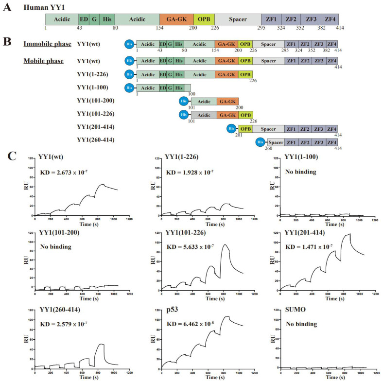
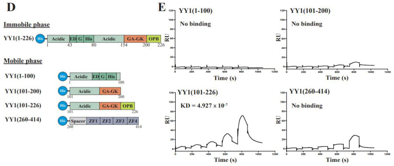
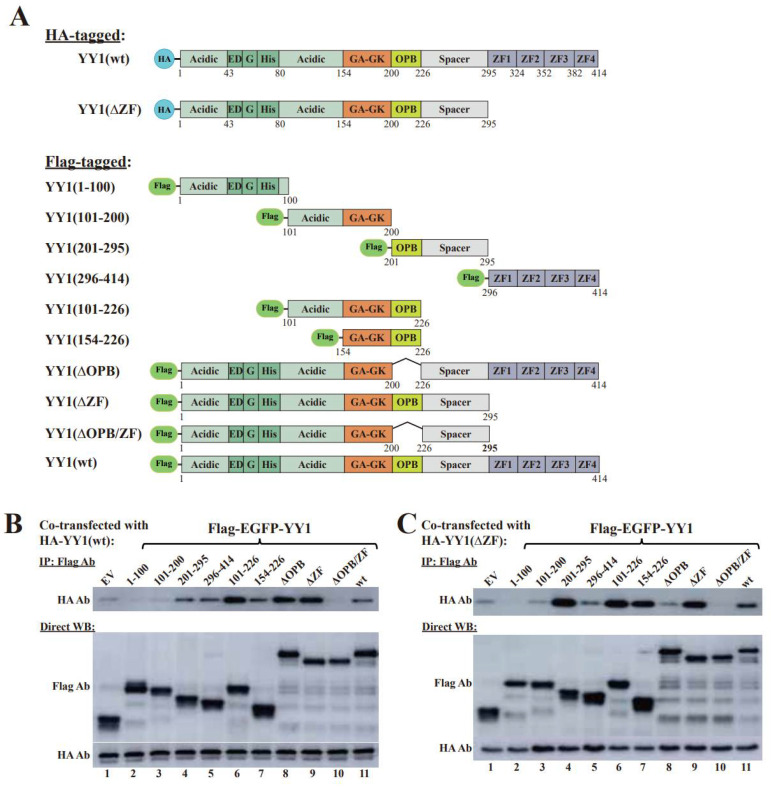
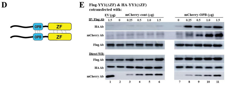
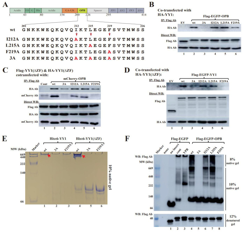
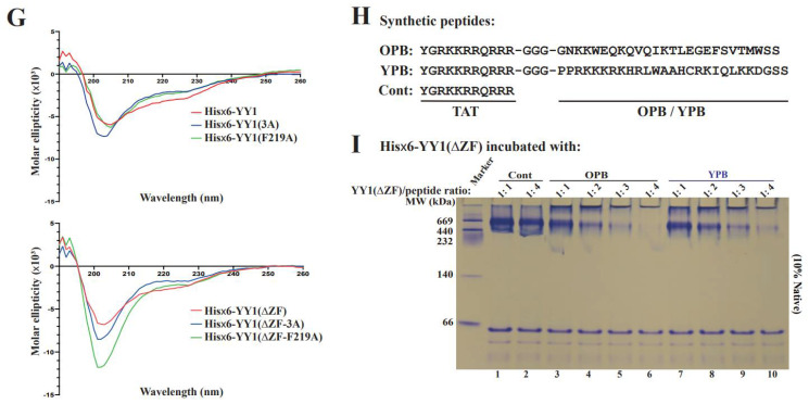
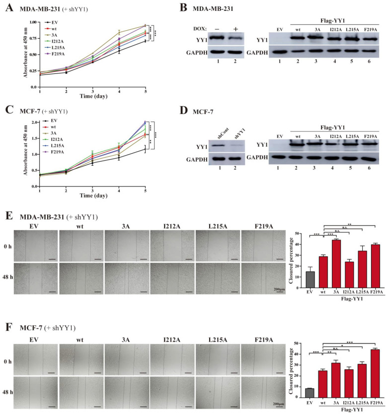
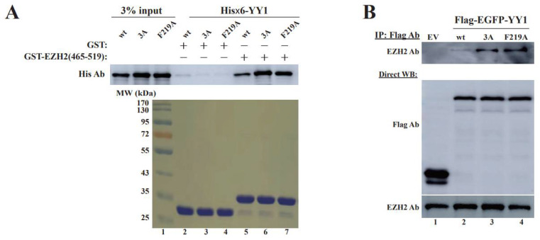
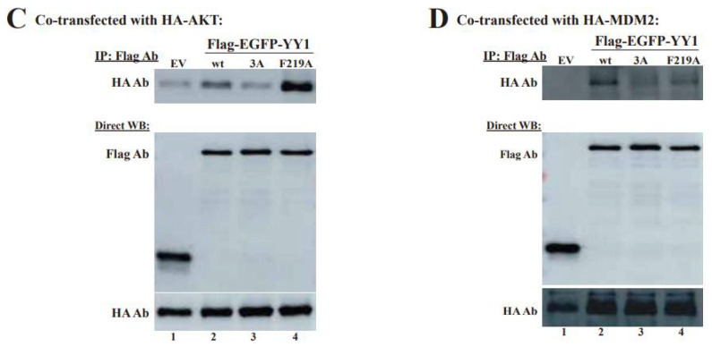
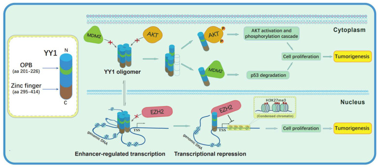
Yin Yang 1 (YY1) plays an oncogenic role through regulating the expression of various cancer-related genes and activating key oncoproteins. Previous research reported that YY1 protein formed dimers or oligomers without definite biological implications. In this study, we first demonstrated the oncoprotein binding (OPB) and zinc finger (ZF) domains of YY1 as the regions involved in its intermolecular interactions. ZFs are well-known for protein dimerization, so we focused on the OPB domain. After mutating three hydrophobic residues in the OPB to alanines, we discovered that YY1(F219A) and YY1(3A), three residues simultaneously replaced by alanines, were defective of intermolecular interaction. Meanwhile, the OPB peptide could robustly facilitate YY1 protein oligomerization. When expressed in breast cancer cells with concurrent endogenous YY1 knockdown, YY1(F219A) and (3A) mutants showed better capacity than wt in promoting cell proliferation and migration, while their interactions with EZH2, AKT and MDM2 showed differential alterations, especially with improved EZH2 binding affinity. Our study revealed a crucial role of the OPB domain in facilitating YY1 oligomerization and suggested a mutually exclusive regulation between YY1-mediated enhancer formation and its activities in promoting oncoproteins.

Keywords: OPB; YY1; oligomerization; oncoprotein; transcription factor; zinc finger.


Conflict of interest statement

The authors declare no competing interest.

Figures

Figure 1
Figure 1
Mapping YY1 binding domains involved in its intermolecular interactions by the surface plasmon resonance (SPR) analysis. (A) Schematic diagrams of the domain structures of YY1 protein. “Acidic” represents a region enriched with aspartic and glutamic acids, and “ED” and “G” depict regions containing a glutamic/aspartic acid cluster and a glycine cluster, respectively. “His” indicates a region consisting of 11 consecutive histidines, and “GA” and “GK” represent glycine/alanine- and glycine/lysine-enriched regions, respectively; OPB: oncoprotein binding; the Spacer region and each C2H2-type Zinc finger domain are also denoted. (B,D) Diagrams of His-tagged (×6) YY1 wt and mutants used to map domains responsible for YY1 intermolecular interactions. (C,E) Evaluation of YY1 intermolecular interactions using the SPR analysis. As the immobile phase, purified His×6-YY1 wt (C) and its mutant (1–226) (E) were individually conjugated to the CM5 chip (GE healthcare). As the mobile phase, purified His×6-YY1 wt and its mutant proteins were serially diluted into concentrations of 62.5, 125, 250, 500 and 1000 nM for injection. The samples individually flowed over the chip channels with different conjugates, and the response units (RU) were received from each single cycle. The binding kinetics were analyzed with the Biacore T200 Evaluation software, version 2.0. The results are displayed with time (s) versus and the RU values.
Figure 1
Figure 1
Mapping YY1 binding domains involved in its intermolecular interactions by the surface plasmon resonance (SPR) analysis. (A) Schematic diagrams of the domain structures of YY1 protein. “Acidic” represents a region enriched with aspartic and glutamic acids, and “ED” and “G” depict regions containing a glutamic/aspartic acid cluster and a glycine cluster, respectively. “His” indicates a region consisting of 11 consecutive histidines, and “GA” and “GK” represent glycine/alanine- and glycine/lysine-enriched regions, respectively; OPB: oncoprotein binding; the Spacer region and each C2H2-type Zinc finger domain are also denoted. (B,D) Diagrams of His-tagged (×6) YY1 wt and mutants used to map domains responsible for YY1 intermolecular interactions. (C,E) Evaluation of YY1 intermolecular interactions using the SPR analysis. As the immobile phase, purified His×6-YY1 wt (C) and its mutant (1–226) (E) were individually conjugated to the CM5 chip (GE healthcare). As the mobile phase, purified His×6-YY1 wt and its mutant proteins were serially diluted into concentrations of 62.5, 125, 250, 500 and 1000 nM for injection. The samples individually flowed over the chip channels with different conjugates, and the response units (RU) were received from each single cycle. The binding kinetics were analyzed with the Biacore T200 Evaluation software, version 2.0. The results are displayed with time (s) versus and the RU values.
Figure 2
Figure 2
Identification of the binding sites responsible for YY1 dimerization by co-immunoprecipitation (co-IP). (A) Diagrams of HA- and Flag-tagged YY1 wt and its mutant proteins used for co-IP studies. (B,C) Co-IP studies to determine the binding sites responsible for YY1 dimerization. HA-YY1 wt (B) or its ΔZF mutant (C) expression plasmid was individually cotransfected with Flag-EGFP-YY1 wt and mutant expression vectors, as well as an empty vector (EV). Cell lysates were co-IPed by a Flag antibody, and HA and Flag antibodies were used in Western blot analyses. (D) Schematic diagram of a predicted YY1 dimerization mode. (E) Effects of the OPB on intermolecular interaction of YY1. Different amounts of mCherry-cont, mCherry-OPB and mCherry plasmids were individually cotransfected with Flag-EGFP-YY1(ΔZF) and HA-YY1(ΔZF) expression vectors, followed by co-IP of the Flag antibody. HA, mCherry and Flag antibodies were used in Western blot analyses.
Figure 2
Figure 2
Identification of the binding sites responsible for YY1 dimerization by co-immunoprecipitation (co-IP). (A) Diagrams of HA- and Flag-tagged YY1 wt and its mutant proteins used for co-IP studies. (B,C) Co-IP studies to determine the binding sites responsible for YY1 dimerization. HA-YY1 wt (B) or its ΔZF mutant (C) expression plasmid was individually cotransfected with Flag-EGFP-YY1 wt and mutant expression vectors, as well as an empty vector (EV). Cell lysates were co-IPed by a Flag antibody, and HA and Flag antibodies were used in Western blot analyses. (D) Schematic diagram of a predicted YY1 dimerization mode. (E) Effects of the OPB on intermolecular interaction of YY1. Different amounts of mCherry-cont, mCherry-OPB and mCherry plasmids were individually cotransfected with Flag-EGFP-YY1(ΔZF) and HA-YY1(ΔZF) expression vectors, followed by co-IP of the Flag antibody. HA, mCherry and Flag antibodies were used in Western blot analyses.
Figure 3
Figure 3
Characterization of key amino acids in charge of YY1 intermolecular interaction. (A) Diagrams of OPB point mutations to determine key hydrophobic amino acids involved in intermolecular interactions among YY1 molecules. (B) Evaluation of YY1 interaction with OPB wt and mutants. Plasmids of Flag-EGFP-OPB wt and mutants were individually cotransfected with the HA-YY1 expression vector, followed by IP of the Flag antibody and Western blot analyses using labeled antibodies. (C) Examination of the effects of OPB wt and mutants on YY1 intermolecular interaction. Plasmids of mCherry-OPB wt, mutants, and cont were individually cotransfected with Flag-YY1(ΔZF) and HA-YY1(ΔZF), followed by IP using the Flag antibody and Western blot analyses using labeled antibodies. (D) Evaluation of the interaction between YY1 and YY1 with wt or mutated OPB sequence. Plasmids of Flag-EGFP-YY1(wt) and OPB mutants were individually cotransfected with HA-YY1(ΔZF), followed by IP using the Flag antibody and Western blot analysis using labeled antibodies. (E) Native polyacrylamide gel electrophoresis (PAGE) to evaluate the effects of OPB wt and mutants on YY1 intermolecular interactions. Purified His×6-YY1(wt) and (ΔZF) proteins with OPB of wt, 3A or F219A sequences were analyzed by a 10% native PAGE. The red arrow heads denote the oligomerization of YY1 proteins. (F) IP studies to examine the binding protein patterns by OPB wt and mutants. HeLa cells were individually transfected by Flag-EGFP-OPB wt, different OPB mutants, YPB, cont and no insert vectors, followed by IP using the Flag antibody, resolved by native gels or SDS-containing gel and analyzed by Western blot using the Flag antibody. (G) Circular dichroism spectroscopy of purified YY1 and its mutants. Purified His×6-YY1(wt) and (ΔZF) proteins (0.05 mg/mL) with wt or mutated OPB domain were individually scanned between the wavelengths from 190 to 280 nm at 20 °C. The data were analyzed by the GraphPad Prism 5.0 software. (H) The sequences of the synthetic OPB, YPB and Cont peptides. TAT: a cell-penetrating peptide (CPP) derived from human immunodeficiency virus. (I) Examination of the effects of OPB and YPB peptides on YY1 oligomerization. Purified His×6-YY1(ΔZF) protein (6.5 µg, or 140 pmol) was mixed with synthetic OPB, YPB and Cont peptides with a serial molar ratio of 1:1, 1:2, 1:3 and 1:4, and incubated at 4 °C for 2 h, followed by the analysis of 10% native PAGE.
Figure 3
Figure 3
Characterization of key amino acids in charge of YY1 intermolecular interaction. (A) Diagrams of OPB point mutations to determine key hydrophobic amino acids involved in intermolecular interactions among YY1 molecules. (B) Evaluation of YY1 interaction with OPB wt and mutants. Plasmids of Flag-EGFP-OPB wt and mutants were individually cotransfected with the HA-YY1 expression vector, followed by IP of the Flag antibody and Western blot analyses using labeled antibodies. (C) Examination of the effects of OPB wt and mutants on YY1 intermolecular interaction. Plasmids of mCherry-OPB wt, mutants, and cont were individually cotransfected with Flag-YY1(ΔZF) and HA-YY1(ΔZF), followed by IP using the Flag antibody and Western blot analyses using labeled antibodies. (D) Evaluation of the interaction between YY1 and YY1 with wt or mutated OPB sequence. Plasmids of Flag-EGFP-YY1(wt) and OPB mutants were individually cotransfected with HA-YY1(ΔZF), followed by IP using the Flag antibody and Western blot analysis using labeled antibodies. (E) Native polyacrylamide gel electrophoresis (PAGE) to evaluate the effects of OPB wt and mutants on YY1 intermolecular interactions. Purified His×6-YY1(wt) and (ΔZF) proteins with OPB of wt, 3A or F219A sequences were analyzed by a 10% native PAGE. The red arrow heads denote the oligomerization of YY1 proteins. (F) IP studies to examine the binding protein patterns by OPB wt and mutants. HeLa cells were individually transfected by Flag-EGFP-OPB wt, different OPB mutants, YPB, cont and no insert vectors, followed by IP using the Flag antibody, resolved by native gels or SDS-containing gel and analyzed by Western blot using the Flag antibody. (G) Circular dichroism spectroscopy of purified YY1 and its mutants. Purified His×6-YY1(wt) and (ΔZF) proteins (0.05 mg/mL) with wt or mutated OPB domain were individually scanned between the wavelengths from 190 to 280 nm at 20 °C. The data were analyzed by the GraphPad Prism 5.0 software. (H) The sequences of the synthetic OPB, YPB and Cont peptides. TAT: a cell-penetrating peptide (CPP) derived from human immunodeficiency virus. (I) Examination of the effects of OPB and YPB peptides on YY1 oligomerization. Purified His×6-YY1(ΔZF) protein (6.5 µg, or 140 pmol) was mixed with synthetic OPB, YPB and Cont peptides with a serial molar ratio of 1:1, 1:2, 1:3 and 1:4, and incubated at 4 °C for 2 h, followed by the analysis of 10% native PAGE.
Figure 4
Figure 4
Effects of the OPB mutations on cell proliferation and migration of breast cancer cells. (AD) Effects of ectopic expression of Flag-YY1 wt and its OPB mutants on the proliferation of breast cancer cells. MDA-MB-231 cells carrying a TET-inducible shYY1 targeting the YY1 mRNA 3′-UTR cultured in doxycycline (DOX)-containing medium (A), or MCF-7 cells infected by lentivirus carrying the same shYY1 (C), were infected by lentivirus expressing Flag-YY1 wt and mutants, followed by WST-1 assay, to determine cell proliferation. Endogenous YY1 knockdown and ectopic expression of YY1 wt and mutants in MDA-MB-231 and MCF-7 cells are shown in (B,D), respectively. (E,F) Scratch assays to test the effects of YY1 mutants on breast cell migration. Endogenous YY1 knockdown and ectopic YY1 expression in MDA-MB-231 (E) and MCF-7 (F) cells carried out as described in (AD). Images were captured when scratches were just made on the plates of overnight cultured cells and after 48 h of culture in the presence of 1.0 µM of Mitomycin C. The quantitation of cell migration is shown at the right panels. Data represent the mean ± S.D., n.s.: no significance, * p < 0.05, ** p < 0.01 and *** p < 0.001.
Figure 5
Figure 5
Evaluation of the effects of OPB mutations on YY1 interaction with oncoproteins. (A,B) Examination of the interaction between YY1 OPB mutants and EZH2. In A, GST and GST-EZH2(465–519) were individually incubated with purified His×6-YY1 wt, 3A and F219A mutants, followed by extensive wash and Western blot analyses using a His-tag antibody. The input of GST and GST-EZH2(465–519) is shown at the lower panel. In (B), plasmids of Flag-EGFP-YY1 wt, 3A and F219A were individually transfected into HeLa cells, followed by IP using the Flag antibody and Western blot analyses using an EZH2 antibody. Direct Western blot analyses were conducted using the antibodies as labeled. (C,D) Examination of the interaction of YY1 OPB mutants with AKT and MDM2. Flag-EGFP-YY1 plasmid was cotransfected with HA-AKT (C) or HA-MDM2 (D) into HeLa cells, followed by IP using the Flag antibody and Western blot analyses using an HA antibody. Direct Western blot analyses were conducted using the antibodies as labeled. Original images of all Western blot data are available in File S1.
Figure 5
Figure 5
Evaluation of the effects of OPB mutations on YY1 interaction with oncoproteins. (A,B) Examination of the interaction between YY1 OPB mutants and EZH2. In A, GST and GST-EZH2(465–519) were individually incubated with purified His×6-YY1 wt, 3A and F219A mutants, followed by extensive wash and Western blot analyses using a His-tag antibody. The input of GST and GST-EZH2(465–519) is shown at the lower panel. In (B), plasmids of Flag-EGFP-YY1 wt, 3A and F219A were individually transfected into HeLa cells, followed by IP using the Flag antibody and Western blot analyses using an EZH2 antibody. Direct Western blot analyses were conducted using the antibodies as labeled. (C,D) Examination of the interaction of YY1 OPB mutants with AKT and MDM2. Flag-EGFP-YY1 plasmid was cotransfected with HA-AKT (C) or HA-MDM2 (D) into HeLa cells, followed by IP using the Flag antibody and Western blot analyses using an HA antibody. Direct Western blot analyses were conducted using the antibodies as labeled. Original images of all Western blot data are available in File S1.
Figure 6
Figure 6
A schematic model of competitive processes between YY1 oligomerization and its interactions with different oncoproteins. In the nucleus, YY1 oligomers may activate target gene expression through facilitating enhancer formation, but may not be able to bind the oncoproteins. As monomers, YY1 interacts with EZH2, AKT and MDM2 in either the nucleus or cytoplasm to promote their oncogenic activities, including suppressing tumor suppressive genes, stimulating proliferative signaling pathways, and enhancing p53 ubiquitination and degradation, respectively.

Similar articles

Cited by

References

    1. Zhang Q., Stovall D.B., Inoue K., Sui G. The oncogenic role of Yin Yang 1. Crit. Rev. Oncog. 2011;16:163–197. doi: 10.1615/CritRevOncog.v16.i3-4.30. - DOI - PMC - PubMed
    1. Caretti G., Di Padova M., Micales B., Lyons G.E., Sartorelli V. The Polycomb Ezh2 methyltransferase regulates muscle gene expression and skeletal muscle differentiation. Genes Dev. 2004;18:2627–2638. doi: 10.1101/gad.1241904. - DOI - PMC - PubMed
    1. Ye K. Nucleophosmin/B23, a multifunctional protein that can regulate apoptosis. Cancer Biol. Ther. 2005;4:918–923. doi: 10.4161/cbt.4.9.2072. - DOI - PubMed
    1. Glenn D.J., Wang F., Chen S., Nishimoto M., Gardner D.G. Endothelin-stimulated human B-type natriuretic peptide gene expression is mediated by Yin Yang 1 in association with histone deacetylase 2. Hypertension. 2009;53:549–555. doi: 10.1161/HYPERTENSIONAHA.108.125088. - DOI - PMC - PubMed
    1. Sankar N., Baluchamy S., Kadeppagari R.K., Singhal G., Weitzman S., Thimmapaya B. p300 provides a corepressor function by cooperating with YY1 and HDAC3 to repress c-Myc. Oncogene. 2008;27:5717–5728. doi: 10.1038/onc.2008.181. - DOI - PubMed