. 2022 Mar 31;14(7):1773.
doi: 10.3390/cancers14071773.

Inhibition of MACC1-Induced Metastasis in Esophageal and Gastric Adenocarcinomas

Affiliations

Inhibition of MACC1-Induced Metastasis in Esophageal and Gastric Adenocarcinomas

Christoph Treese et al. Cancers (Basel). .

Abstract

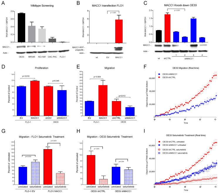
Esophageal and Gastric Adenocarcinomas (AGE/S) are characterized by early metastasis and poor survival. MACC1 (Metastasis Associated in Colon Cancer 1) acts in colon cancer as a metastasis inducer and is linked to reduced survival. This project illuminates the role and potential for the inhibition of MACC1 in AGE/S. Using 266 of 360 TMAs and survival data of AGE/S patients, we confirm the value of MACC1 as an independent negative prognostic marker in AGE/S patients. MACC1 gene expression is correlated with survival and morphological characteristics. In vitro analysis of lentivirally MACC1-manipulated subclones of FLO-1 and OE33 showed enhanced migration induced by MACC1 in both cell line models, which could be inhibited by the MEK1 inhibitor selumetinib. In vivo, the efficacy of selumetinib on tumor growths and metastases of MACC1-overexpressing FLO-1 cells xenografted intrasplenically in NOG mice was tested. Mice with high-MACC1-expressing cells developed faster and larger distant metastases. Treatment with selumetinib led to a significant reduction in metastasis exclusively in the MACC1-positive xenografts. MACC1 is an enhancer of tumor aggressiveness and a predictor of poor survival in AGE/S. This effect can be inhibited by selumetinib.

Keywords: MACC1; MEK1 inhibition; esophageal cancer; gastric cancer; metastasis; selumetinib.


Conflict of interest statement

The authors declare no conflict of interest.

Figures

Figure 1
Figure 1
Representative MACC1 IHC staining of TMA cores. (A) positive staining (100× and 400× magnitude), (B) negative staining (100× and 400× magnitude).
Figure 2
Figure 2
Kaplan–Meier plots of survival, MACC1-low: blue and MACC1-high: red. Significance calculated by log-rank test. (A) Overall survival: MACC1-low, better survival, p = 0.002; (B) Disease specific survival: MACC-low better survival, p = 0.011. (C) Overall survival—subgroup impact of vein invasion. No vein invasion: blue and red as above; MACC-low, better survival, p < 0.001. With vein invasion: MACC1-low: green; MACC1-high: grey; difference not significant, p = 0.637. (D) Overall survival—subgroup impact of lymphatic vessel invasion. No lymphatic vessel invasion: blue and red as above; MACC-low, better survival, p = 0.003; With lymphatic vessel invasion: MACC1-low: green; MACC1-high: grey; difference not significant: p = 0.287.
Figure 3
Figure 3
(A) Western blot and RNA expression of cell lines OE33, MKN45, NCI-N87, OAC-P4C and FLO-1: high MACC1 expression in OE33 cells, negligible MACC1 expression in FLO-1 cells. (B) Western blot and RNA expression of MACC1-transfected cell line FLO-1. wt = wild type, EV = empty vector and MACC1; Significant overexpression of MACC1 in FLO-1/MACC1 compared to FLO-1/EV (p < 0.001). (C) Western blot and RNA expression of different MACC1-downregulated OE33 subclones. wt = wild type, shCTRL = scrambled sh control, shMACC1 1–4 = 4 different MACC1 small hairpin vectors. (D) The FLO-1/MACC1 clone shows higher proliferation over 24 h compared to FLO-1/EV (p = 0.010). OE33/shMACC1 showed no statistically significant difference (p = 0.345) in comparison to OE33/shCTRL. (E) In the migration assay, over 72 h FLO-1/MACC1 cells migrate significantly more than FLO-1/EV cells (p = 0.004), and OE33/shMACC1 cells significantly less than OE33/shCTRL cells (p = 0.013). (F) Effects of MACC1 inhibition on migration in a real time plot over 72 h for OE33/shMACC1-negative (blue) and MACC1-positive OE33/shCTRL (red) cells. (G) Treatment with selumetinib (light colors) decreases migration of FLO-1/MACC1 cells (red) compared to untreated cells (dark colors) significantly (p < 0.001). (H) Treatment with selumetinib (light colors) decreases migration of MACC1-positive OE33/shCTRL cells significantly compared to migration in untreated cells (p = 0.001) (red). (I) Effects on migration by inhibition with selumetinib in a real time plot over 72 h: MACC1 knockdown OE33/shMACC1 cells in blue and MACC1-positive OE33/shCTRL in red. Selumetinib-treated cells are shown in light and untreated in dark colors. Significant effects on selumetinib treatment are only shown in the MACC1-expressing OE33/shCTRL cells.
Figure 4
Figure 4
Effect of selumetinib (AZD6244) on MACC1-induced metastasis formation in a mouse xenograft model. (A) NOG mice were xenotransplanted with FLO-1/EV/Luc and FLO-1/MACC1/Luc cells by intrasplenal injection (n = 6 per group). Metastasis formation in the liver was monitored by BLI for 21 days (left panel). Representative BLI overlays of the animals are shown in the right panel. In the next experiment, NOG mice (n = 10 per group) were xenotransplanted with FLO-1/EV/Luc and FLO-1/MACC/Luc. Mice were treated with either vehicle or AZD6244. (BD) Metastasis formation in the liver was analyzed after three weeks by BLI (B) and human satellite DNA (C), and visualized by human CK19 staining of murine liver tissue (D). *: p < 0.05.

References

    1. Sung H., Ferlay J., Siegel R.L., Laversanne M., Soerjomataram I., Jemal A., Bray F. Global Cancer Statistics 2020: GLOBOCAN Estimates of Incidence and Mortality Worldwide for 36 Cancers in 185 Countries. CA Cancer J. Clin. 2021;71:209–249. doi: 10.3322/caac.21660. - DOI - PubMed
    1. Son T. Multi-institutional validation of the 8th AJCC TNM staging system for gastric cancer: Analysis of survival data from high-volume Eastern centers and the SEER database. J. Surg. Oncol. 2019;120:676–684. doi: 10.1002/jso.25639. - DOI - PubMed
    1. Riihimäki M., Hemminki A., Sundquist K., Sundquist J., Hemminki K. Metastatic spread in patients with gastric cancer. Oncotarget. 2016;7:52307–52316. doi: 10.18632/oncotarget.10740. - DOI - PMC - PubMed
    1. He X., Wu W., Lin Z., Ding Y., Si J., Sun L. Validation of the American Joint Committee on Cancer (AJCC) 8th edition stage system for gastric cancer patients: A population-based analysis American Joint Committee on Cancer. Gastric Cancer. 2018;21:391–400. doi: 10.1007/s10120-017-0770-1. - DOI - PubMed
    1. Stein U., Walther W., Arlt F., Schwabe H., Smith J., Fichtner I., Birchmeier W., Schlag P.M. MACC1, a newly identified key regulator of HGF-MET signaling, predicts colon cancer metastasis. Nat. Med. 2009;15:59–67. doi: 10.1038/nm.1889. - DOI - PubMed