Review
. 2022 Apr 6;14(7):1835.
doi: 10.3390/cancers14071835.

Neuroendocrine Neoplasms of the Gynecologic Tract

Affiliations
Review

Neuroendocrine Neoplasms of the Gynecologic Tract

Mayur Virarkar et al. Cancers (Basel). .

Abstract

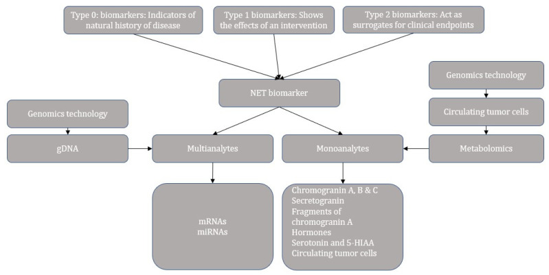
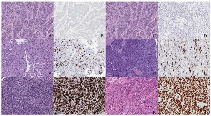
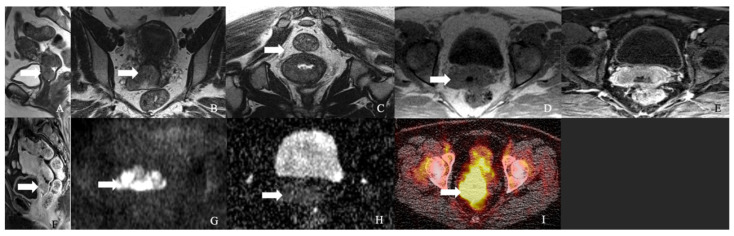
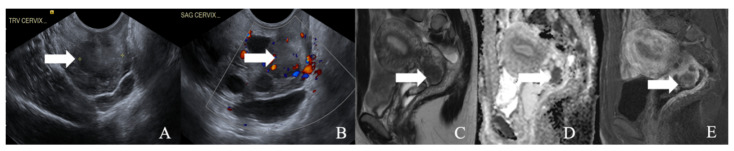
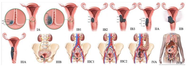
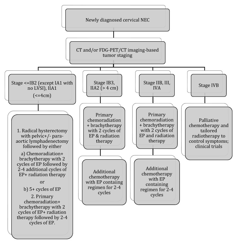
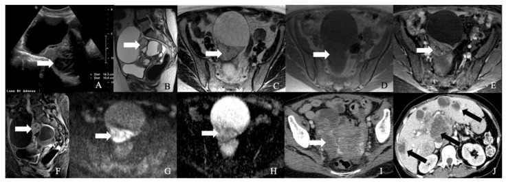
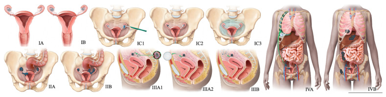
Gynecological tract neuroendocrine neoplasms (NEN) are rare, aggressive tumors from endocrine cells derived from the neuroectoderm, neural crest, and endoderm. The primary gynecologic NENs constitute 2% of gynecologic malignancies, and the cervix is the most common site of NEN in the gynecologic tract. The updated WHO classification of gynecologic NEN is based on the Ki-67 index, mitotic index, and tumor characteristics such as necrosis, and brings more uniformity in the terminology of NENs like other disease sites. Imaging plays a crucial role in the staging, triaging, restaging, and surveillance of NENs. The expression of the somatostatin receptors on the surface of neuroendocrine cells forms the basis of increasing evaluation with functional imaging modalities using traditional and new tracers, including 68Ga-DOTA-Somatostatin Analog-PET/CT. Management of NENs involves a multidisciplinary approach. New targeted therapies could improve the paradigm of care for these rare malignancies. This article focuses on the updated staging classifications, clinicopathological characteristics, imaging, and management of gynecologic NENs of the cervix, ovary, endometrium, vagina, and vulva, emphasizing the relatively common cervical neuroendocrine carcinomas among these entities.

Keywords: FIGO classification; PET/CT; cervical neuroendocrine tumor; gynecological NENs; imaging of neuroendocrine tumors; neuroendocrine.


Conflict of interest statement

The authors declare no conflict of interest.

Figures

Figure 1
Figure 1
Categories and progress overview of neuroendocrine tumor biomarkers.
Figure 2
Figure 2
(A,B) Well-differentiated NET grade 1. (A) Well-differentiated neuroendocrine tumor grade 1 (low grade) with an organoid pattern, with a meshwork of thin fibrovascular septa surrounding nests of tumor cells. Tumor cells are uniform with a polygonal shape, round to oval nuclei with salt and pepper chromatin, inconspicuous nucleoli, and moderate to abundant eosinophilic cytoplasm. (B) The Ki67 immunohistochemical stain shows a proliferation rate of 2%. (C,D) Well-differentiated NET grade 2. (C) Neuroendocrine cells in a well-differentiated neuroendocrine tumor, grade 2. Tumor cells are relatively uniform and round. The nuclear chromatin is finely granular. (D) The Ki67 immunohistochemical stain shows a proliferation rate of 10%. (E,F) Well-differentiated NET grade 3. (E) Neuroendocrine cells in a well-differentiated neuroendocrine tumor, grade 3. Tumor cells are relatively uniform and round with eosinophilic cytoplasm. The nuclear chromatin is granular. (F) The Ki67 immunohistochemical stain shows a proliferation rate of 30–40%. (G,H) Poorly differentiated neuroendocrine carcinoma. (G) The tumor shows solid nests of poorly differentiated epithelioid cells with dense chromatin. (H) Ki67 immunohistochemical stain shows a proliferation rate of 80%. (I,J) Small cell carcinoma. (I) Sheets of oval blue cells with minimal cytoplasm. The chromatin is dense. Nuclei demonstrate molding and smudging. (J) Ki67 shows a proliferation index of 80%. (K,L) Large cell neuroendocrine carcinoma. (K) Tumor cells with sheets of large, epithelioid cells. Cytologic features show abundant cytoplasm, coarse chromatin, nuclear pleomorphism, and prominent nucleoli. (L) Ki67 is greater than 90%.
Figure 3
Figure 3
A 62-year-old female with neuroendocrine carcinoma of the cervix. (A) Sagittal T2 weighted image, (B) axial T2 weighted image, (C) oblique T2 weighted image, (D) axial pre-contrast T1 weighted image, (E) axial fat-saturated post-contrast T1 weighted image, (F) sagittal fat-saturated post-contrast T1 weighted image, (G) axial diffusion-weighted images (B-100), (H) axial apparent diffusion coefficient MRI images, and (I) Axial Ga-68 dotatate PET/CT image demonstrate a FDG avid enhancing mass in the cervix (arrow) with restricted diffusion and no parametrium involvement. The mass biopsy reported a small cell neuroendocrine carcinoma.
Figure 4
Figure 4
A 52-year-old female with a neuroendocrine tumor of the cervix uteri. (A) Transverse and (B) sagittal ultrasound image of the cervix demonstrates a heterogenous cervical mass (arrow) measuring about 14 cm. (C) Sagittal T2 weighted image, (D) sagittal apparent diffusion coefficient map, and (E) sagittal fat-saturated post-contrast T1 weighted MRI images demonstrate a mass in the cervix uteri (arrow) with restricted diffusion. The mass biopsy reported a small cell neuroendocrine tumor of grade G3.
Figure 5
Figure 5
2018 FIGO staging system for cervical cancer. Stage I, confined to the cervix. Stage IA, ≤5 mm depth. Stage IA1, ≤3 mm depth. Stage IA2, 3 mm and ≤5 mm depth. Stage IB, >5 mm depth. Stage IB1, ≤2 cm maximum diameter. Stage IB2, >2 cm and ≤4 cm maximum diameter. Stage IB3, >4 cm maximum diameter. Stage II, beyond the uterus but not involving the lower one-third of the vagina or pelvic sidewall. Stage IIA, upper two-thirds of the vagina. Stage IIA1, upper two-thirds of the vagina and ≤4 cm. Stage IIA2, Upper two-thirds of the vagina and >4 cm. Stage IIB, parametrial invasion. Stage III, lower vagina, pelvic sidewall, ureters, and lymph nodes. Stage IIIA, lower one-third of the vagina. Stage IIIB, pelvic sidewall. Stage IIIC, pelvic, and para-aortic lymph node involvement. Stage IIIC1, pelvic lymph node involvement. Stage IIIC2, para-aortic lymph node involvement. Stage IV, adjacent and distant organs. Stage IVA, rectal or bladder involvement. Stage IVB, distant organs outside the pelvis.
Figure 6
Figure 6
Stage-based treatment algorithm for cervical neuroendocrine carcinoma [2]. NEC: neuroendocrine carcinoma; LVSI: lymphovascular space invasion; EP: etoposide and cisplatin; chemotherapy with EP alone: cisplatin 60–80 mg/m2 on day 1 every 3 weeks and etoposide 80–120 mg/m2 on days 1–3 every 3 weeks (with growth factor support); chemotherapy with EP + radiation therapy: 2 cycles of EP q3 weeks (cisplatin 60 mg/m2 and etoposide 100 mg/m2 given on day 1 of 21 day cycle) during radiation therapy followed by an additional 2–4 cycles of EP alone (recommended regimen). Radiotherapy: 40–45 Gy external beam radiotherapy ±40–45 Gy brachytherapy (modified from Winer et al.).
Figure 7
Figure 7
52-year-old female with a neuroendocrine tumor of the ovary. (A) Transverse ultrasound image of the pelvis demonstrates a heterogeneous mass in the pelvis (arrow) measuring about 14 cm. (B) Sagittal T2 weighted image, (C) axial T2 weighted image, (D) axial pre-contrast T1 weighted image, (E) axial fat-saturated post-contrast T1 weighted image, (F) sagittal fat-saturated post-contrast T1 weighted image, (G) axial diffusion-weighted images (B-100), (H) axial apparent diffusion coefficient images demonstrate a multiloculated mass with enhancing nodule (arrow) and restricted diffusion, (I) axial post-contrast CT image of the pelvis shows the heterogenous mass (arrow), and (J) axial post-contrast CT image of the abdomen demonstrates the multiple hepatic metastases (arrow). The mass was resected, and a biopsy reported neuroendocrine tumor of the ovary with synaptophysin, CD56, CK7, and CDX-2 were positive, and chromogranin was focally positive, with a Ki67 index of less than 5%.
Figure 8
Figure 8
FIGO staging system for ovarian cancer. In stage IA cancer, the tumor is limited to one ovary or fallopian tube. The tumor is limited to both ovaries or fallopian tubes in stage IB cancer. Stage IC1 cancer results from the intraoperative spill. In stage IC3 cancer, malignant cells are found in ascites or peritoneal washings. In stage IIA, the tumor extends to or is implanted on (or both) uterus or fallopian tubes (or both). Stage IIIA1 cancer shows positive retroperitoneal lymph nodes. Stage IIIA2 cancer, microscopic, extrapelvic (above pelvic brim) peritoneal involvement is seen with or without positive retroperitoneal lymph nodes. Stage IIIB or stage IIIC cancer, macroscopic, extrapelvic (above pelvic brim) peritoneal involvement is seen with or without positive retroperitoneal lymph nodes (≤2 cm for stage IIIB and >2 cm for stage IIIC). Stage IVA cancer indicates pleural effusion with positive cytology. Stage IVB cancer, hepatic or splenic parenchymal metastasis is seen (or both) and metastasis to extra-abdominal organs.
Figure 9
Figure 9
Diagnostic algorithm for ovarian NENs.
Figure 10
Figure 10
Treatment algorithm of ovarian NEC. If non-surgical or metastatic disease: palliative chemotherapy (EP regimen) or enrollment into clinical trials.
Figure 11
Figure 11
A 52-year-old female with a large cell neuroendocrine tumor of the endometrium. (A) Transverse ultrasound image of the uterus demonstrates thickened endometrium (arrow), (B) axial T2 weighted image, (C) sagittal T2 weighted image, (D) sagittal apparent diffusion coefficient map, and (E) sagittal fat-saturated post-contrast T1 weighted MRI images demonstrate a hypoenhancing tumor in the endometrium (arrow) with restricted diffusion.
Figure 12
Figure 12
2009 FIGO staging system for endometrial cancer. Stage I tumor confined to the uterus. Stage IA < 50% myometrial invasion. Stage IB ≥ 50% myometrial invasion. Stage II Cervical stromal invasion. Stage IIIA tumor invasion into serosa or adnexa. Stage IIIB vaginal or parametrial involvement. Stage IIIC1 pelvic node involvement. Stage IIIC2 paraaortic node involvement. Stage IVA tumor invasion into bladder or bowel mucosa. Stage IVB distant metastases (including abdominal metastases) or inguinal lymph node involvement.
Figure 13
Figure 13
Treatment algorithm of endometrial NEC. If non-surgical or metastatic disease: palliative chemotherapy with EP regimen or enrollment into clinical trials.
Figure 14
Figure 14
FIGO Staging of vaginal tumors. Stage I is limited to the vaginal wall. Stage II is beyond the vaginal wall without pelvic sidewall involvement. Stage III extends to the pelvic sidewall. Stage IVA infiltrates bladder or rectum or the tumor extending beyond the pelvis, with any nodal metastasis. Stage IVB has distant metastasis.
Figure 15
Figure 15
Treatment algorithm of vaginal/vulvar NEC. If non-surgical or metastatic disease: palliative chemotherapy with EP regimen or enrollment in clinical trials.
Figure 16
Figure 16
New 2021 FIGO Staging of vulvar tumors. Stage-I tumor confined to the vulva. Stage IA tumor size less than equal to 2 cm and stromal invasion less than equal to 1 mm. Stage IB tumor size more than 2 cm and stromal invasion more than 1 mm. Stage II tumor of any size with extension to lower one-third of the urethra, lower one-third of the vagina, lower one-third of the anus with negative nodes. Stage III tumor of any size with extension to the upper part of adjacent perineal structures or with any number of nonfixed, nonulcerated lymph nodes. Stage IIIA tumor of any size with disease extension to the upper two-thirds of the urethra, upper two-thirds of the vagina, bladder mucosa, rectal mucosa, or regional lymph node metastases less than equal to 5 mm. Stage IIIB regional lymph node metastases more than 5 mm. Stage IIIC regional lymph node metastases with extracapsular spread. Stage IV tumor of any size fixed to bone or fixed, ulcerated lymph node metastases, or distant metastases. Stage IVA disease fixed to the pelvic bone or fixed or ulcerated regional lymph node metastases. Stage IVB distant metastases.

References

    1. Galgano S.J., Sharbidre K., Morgan D.E. Multimodality imaging of neuroendocrine tumors. Radiol. Clin. 2020;58:1147–1159. doi: 10.1016/j.rcl.2020.07.008. - DOI - PubMed
    1. Dasari A., Shen C., Halperin D., Zhao B., Zhou S., Xu Y., Shih T., Yao J.C. Trends in the incidence, prevalence, and survival outcomes in patients with neuroendocrine tumors in the United States. JAMA Oncol. 2017;3:1335–1342. doi: 10.1001/jamaoncol.2017.0589. - DOI - PMC - PubMed
    1. Migut A.E., Kaur H., Avritscher R. Neuroendocrine tumors: Imaging of treatment and follow-up. Radiol. Clin. 2020;58:1161–1171. doi: 10.1016/j.rcl.2020.08.002. - DOI - PubMed
    1. Heller M.T., Shah A.B. Imaging of neuroendocrine tumors. Radiol. Clin. 2011;49:529–548. doi: 10.1016/j.rcl.2011.02.011. - DOI - PubMed
    1. Tempfer C.B., Tischoff I., Dogan A., Hilal Z., Schultheis B., Kern P., Rezniczek G.A. Neuroendocrine carcinoma of the cervix: A systematic review of the literature. BMC Cancer. 2018;18:530. doi: 10.1186/s12885-018-4447-x. - DOI - PMC - PubMed

LinkOut - more resources