Review
. 2022 Mar 28;11(7):1135.
doi: 10.3390/cells11071135.

Artificial Oocyte: Development and Potential Application

Affiliations
Review

Artificial Oocyte: Development and Potential Application

Reza K Oqani et al. Cells. .

Abstract

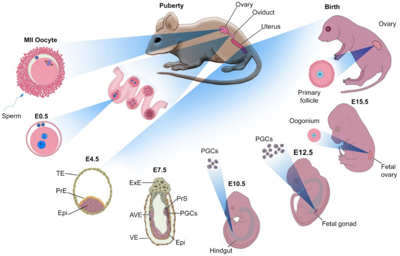
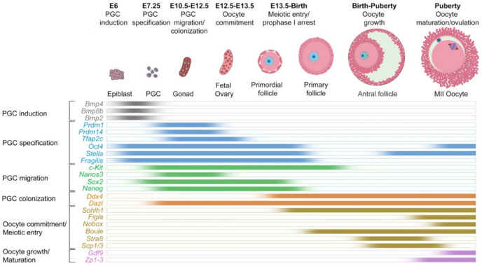
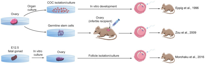
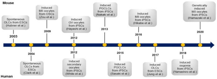
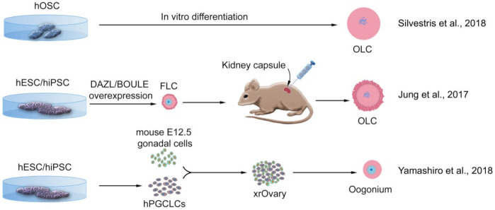
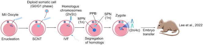
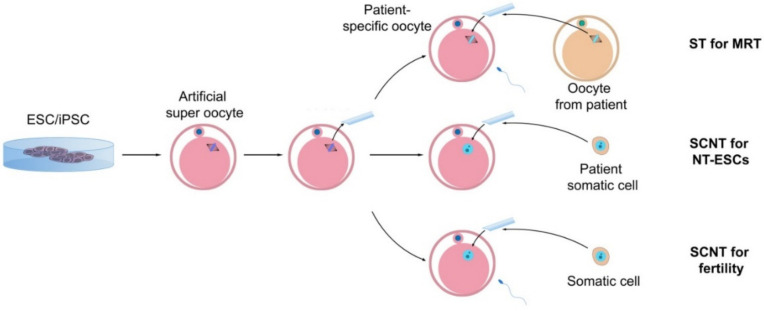
Millions of people around the world suffer from infertility, with the number of infertile couples and individuals increasing every year. Assisted reproductive technologies (ART) have been widely developed in recent years; however, some patients are unable to benefit from these technologies due to their lack of functional germ cells. Therefore, the development of alternative methods seems necessary. One of these methods is to create artificial oocytes. Oocytes can be generated in vitro from the ovary, fetal gonad, germline stem cells (GSCs), ovarian stem cells, or pluripotent stem cells (PSCs). This approach has raised new hopes in both basic research and medical applications. In this article, we looked at the principle of oocyte development, the landmark studies that enhanced our understanding of the cellular and molecular mechanisms that govern oogenesis in vivo, as well as the mechanisms underlying in vitro generation of functional oocytes from different sources of mouse and human stem cells. In addition, we introduced next-generation ART using somatic cells with artificial oocytes. Finally, we provided an overview of the reproductive application of in vitro oogenesis and its use in human fertility.

Keywords: artificial oocyte; assisted reproductive technologies; haploidization of somatic chromosomes; oogenesis; stem cells.


Conflict of interest statement

The authors declare no conflict of interest.

Figures

Figure 1
Figure 1
The development of mouse embryo in vivo. Fertilization, cleavage, and morula compaction take place in the oviduct. On an embryonic day 4.5 (E4.5), the advanced blastocyst is positioned in the uterus and is composed of epiblast (Epi), primitive endoderm (PrE), and trophectoderm (TE). On E7.5, the embryo is composed of extra-embryonic ectoderm (ExE), anterior visceral endoderm, visceral endoderm (VE), Epi, and primitive streak (PrS). At this stage, it has begun gastrulation and is connected to the uterus through the ExE. Several cells in the PrS receive signals from ExE and VE that commit them to differentiate into primordial germ cells (PGCs). With the formation of the gut, PGCs migrate through the hindgut, and on the E12.5, they are colonized and multiply in the gonad (which arises from the genital ridge). With the development of the ovary from the gonad, PGCs go through the early stages of differentiation into the oocyte, stopping shortly after birth in the germinal vesicle (GV) stage. With puberty and secretion of gonadotropins, the development of the oocyte is complete and an MII oocyte is formed. This MII oocyte exits the ovary so that it may enter the oviduct, ready for fertilization.
Figure 2
Figure 2
Expression dynamics of key genes regulating the specification of primordial germ cells and the development of oocytes in vivo in mice.
Figure 3
Figure 3
Successful methods of deriving oocytes from mouse ovarian stem cells. Adult ovaries can be a source of cumulus-oocyte complex (COC) after organ culture, or female germline stem cells (GSCs). The GSCs can differentiate into MII oocytes after transplantation into a sterile recipient’s ovary. Fetal gonads can also develop into ovaries in vitro, and secondary follicles can be isolated and cultured from them, allowing them to develop into MII oocytes. All of these strategies led to producing live offspring [43,44,45].
Figure 4
Figure 4
Landmark studies in mouse and human oocyte differentiation from pluripotent stem cells. Only a few of the most effective studies have been mentioned. These works, along with other efforts discussed in the text, provided a better understanding of how germ cells differentiate in humans and mice. OLCs: oocyte-like cells; ESCs: embryonic stem cells; OSCs: ovarian stem cells; GV: germinal vesicle; iPSCs: induced pluripotent stem cells; PGCLCs: primordial germ cell-like cells.
Figure 5
Figure 5
Successful methods of deriving oocytes in vitro from mouse pluripotent stem cells. Cells originated from mouse embryonic stem cells (mESCs) or mouse-induced pluripotent stem cells (miPSCs) developed into primordial germ cell-like cells (PGCLCs) and differentiated into oocytes either by aggregating with in vivo-derived fetal gonadal somatic cells or with fetal ovarian somatic cell-like cells (FOSLCs), after transplant into the ovarian bursa or by generating reconstituted ovary (rOvary) containing growing oocytes. All of these strategies led to producing live offspring [80,82,85].
Figure 6
Figure 6
Successful methods of deriving oocyte-like cells in vitro from human pluripotent stem cells or ovarian stem cells. Cells that originated from human oogonial stem cells (hOSCs) directly differentiated into oocyte-like cells (OLCs). Human embryonic stem cells (hESCs), or human-induced pluripotent stem cells (hiPSCs), developed into follicle-like cells (FLCs), transplanted into the mouse kidney capsule, and developed to OLCs; or, hESCs or hiPSCs differentiated into primordial germ cell-like cells (PGCLCs), aggregated with mouse fetal gonadal somatic cells, form xenogenic reconstituted ovary (xrOvary) and further differentiated into pre-meiotic oogonium. None of these strategies led to producing meiotic oocytes [90,96,100].
Figure 7
Figure 7
Induction of haploidy in a somatic cell by the mature oocyte. The G0/G1 somatic cell was transferred into the enucleated MII oocyte and the de novo spindle consisting of somatic homologous chromosomes was reconstructed. Fertilization triggered both homologous segregation and extrusion of pseudo polar body (PPB), and further, generation of the somatic pronucleus (SPN) and male pronucleus (MPN). The haploid somatic and sperm pronuclei were formed in the reconstructed zygote. The embryo with the somatic haploid can then develop and produce live offspring. SCNT: somatic cell nuclear transfer [145].
Figure 8
Figure 8
Application of artificial super-oocytes for patient-specific stem cells and infertility treatment. Oocytes can be obtained from pluripotent stem cells, and after enucleating, can be used either for spindle transfer (ST) for mitochondrial replacement therapy (MRT), transfer of the somatic cell nucleus (SCNT) from the patient’s cells for embryonic stem cell (ESC) culture, or nuclear transfer-derived ESCs (NT-ESCs) for fertility treatment and generation of the new oocytes.

References

    1. Bahamondes L., Makuch M.Y. Infertility care and the introduction of new reproductive technologies in poor re-source settings. Reprod. Biol. Endocrinol. 2014;12:1–7. doi: 10.1186/1477-7827-12-87. - DOI - PMC - PubMed
    1. Practice Committee of the American Society for Reproductive Medicine Definitions of infertility and recurrent pregnancy loss: A committee opinion. Fertil. Steril. 2020;113:533–535. doi: 10.1016/j.fertnstert.2019.11.025. - DOI - PubMed
    1. Datta J., Palmer M., Tanton C., Gibson L., Jones K., Macdowall W., Glasier A., Sonnenberg P., Field N., Mercer C., et al. Prevalence of infertility and help seeking among 15,000 women and men. Hum. Reprod. 2016;31:2108–2118. doi: 10.1093/humrep/dew123. - DOI - PMC - PubMed
    1. Hendriks S., Dancet E.A., van Pelt A.M., Hamer G., Repping S. Artificial gametes: A systematic review of biologi-cal progress towards clinical application. Hum. Reprod. Update. 2015;21:285–296. doi: 10.1093/humupd/dmv001. - DOI - PubMed
    1. Chon S.J., Umair Z., Yoon M.-S. Premature Ovarian Insufficiency: Past, Present, and Future. Front. Cell Dev. Biol. 2021;9:672890. doi: 10.3389/fcell.2021.672890. - DOI - PMC - PubMed

Publication types