Review
. 2022 Mar 29;11(7):1155.
doi: 10.3390/cells11071155.

NGLY1 Deficiency, a Congenital Disorder of Deglycosylation: From Disease Gene Function to Pathophysiology

Affiliations
Review

NGLY1 Deficiency, a Congenital Disorder of Deglycosylation: From Disease Gene Function to Pathophysiology

Ashutosh Pandey et al. Cells. .

Abstract

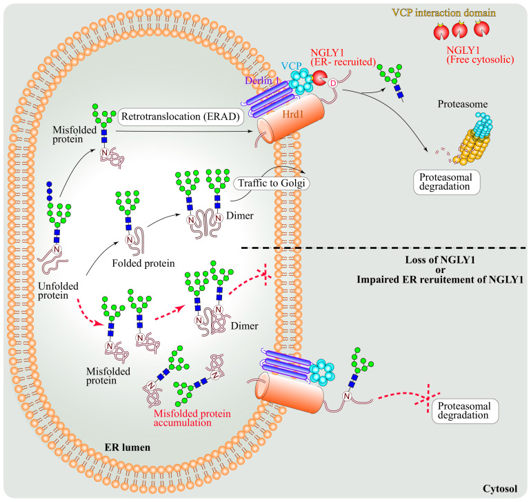
N-Glycanase 1 (NGLY1) is a cytosolic enzyme involved in removing N-linked glycans of misfolded N-glycoproteins and is considered to be a component of endoplasmic reticulum-associated degradation (ERAD). The 2012 identification of recessive NGLY1 mutations in a rare multisystem disorder has led to intense research efforts on the roles of NGLY1 in animal development and physiology, as well as the pathophysiology of NGLY1 deficiency. Here, we present a review of the NGLY1-deficient patient phenotypes, along with insights into the function of this gene from studies in rodent and invertebrate animal models, as well as cell culture and biochemical experiments. We will discuss critical processes affected by the loss of NGLY1, including proteasome bounce-back response, mitochondrial function and homeostasis, and bone morphogenetic protein (BMP) signaling. We will also cover the biologically relevant targets of NGLY1 and the genetic modifiers of NGLY1 deficiency phenotypes in animal models. Together, these discoveries and disease models have provided a number of avenues for preclinical testing of potential therapeutic approaches for this disease.

Keywords: AMPK signaling; BMP signaling; ER-associated degradation (ERAD); N-glycosylation; congenital disorder of deglycosylation (CDDG); deglycosylation; human developmental disorder; mitochondrial abnormality; proteasome; rare disease.


Conflict of interest statement

The authors declare no conflict of interest. The funders had no role in the design of the study, in the writing of the manuscript, or in the decision to publish the manuscript.

Figures

Figure 1
Figure 1
Schematic representation of N-glycanase 1 orthologs with domains present in the indicated species. aa, amino acid; PAW, a domain in PNGases and other worm proteins; PUB, a domain in PNGase/UBA or UBX-containing proteins; TG, transglutaminase-like.
Figure 2
Figure 2
(A) Schematic representation of the reported missense, nonsense, frameshift, and in-frame deletion variants in NGLY1. Nonsense variants are in maroon font above the NGLY1 schematic. Missense (black font), frameshift (blue font), and in-frame deletion (red font) are shown below NGLY1. Protein structure is based on SMART (simple molecular architecture research tool; http://smart.embl-heidelberg.de accessed on 21 March 2022). (B) Ring chart diagram showing the reported variants, including splice site variants (green font) with their relative occurrence. Note that R401X is the most common variant (~21%). aa, amino acid; fs, frameshift; PAW, a domain in PNGases and other worm proteins; PUB, a domain in PNGase/UBA or UBX-containing proteins; TG, transglutaminase-like.
Figure 3
Figure 3
Schematic representation of the cap ’n’ collar proteins in the indicated species. Note that only representative isoforms of the fly cap ’n’ collar are shown. aa, amino acid; NTD, N-terminal domain; NST, Asn/Ser/Thr-rich domain; CNC, cap ’n’ collar; bZIP, basic leucine zipper; BR, basic region.
Figure 4
Figure 4
Schematic representation showing that under normal conditions, retrotranslocation from ER to cytosol directs NFE2L1 to its proteasomal degradation. Upon proteasomal inhibition, NFE2L1 undergoes de-N-glycosylation by NGLY1 and is cleaved by an aspartic protease called DNA damage-inducible 1 homolog 2 (DDI2), which leads to its activation. Activated NFE2L1 enters the nucleus and induces the transcription of proteasomal subunit genes, as well as its other target genes. N, asparagine; D, aspartic acid.
Figure 5
Figure 5
Schematic representation showing poor gut contraction in Pngl-mutant Drosophila midgut accompanied by reduced AMP-Activated Protein Kinase α (AMPKα) levels, increased AMP/ATP ratio, increased reactive oxygen species (ROS) levels, and mitochondrial anomaly. The cartoon also shows the AMPK activators metformin, PT1, and AICAR, as well as the possible mechanisms for regulation of AMPKα mRNA levels by NGLY1. NGLY1 might affect AMPKα transcription by altering specific upstream N-glycosylated proteins. In the absence of NGLY1, ENGASE may result in the accumulation and aggregation of proteins with N-GlcNAc residues, which might interfere with the function of proteins harboring O-GlcNAc. “?” marks potential mechanisms for the Regulation of AMPKα mRNA level by NGLY1.
Figure 6
Figure 6
Schematic representation showing that de-N-glycosylation of misfolded bone morphogenetic protein 4 (BMP4) by endoplasmic reticulum (ER) membrane-recruited NGLY1 promotes its retrotranslocation from ER to cytosol, where it undergoes proteasomal degradation. In the absence of NGLY1 or its impaired ER recruitment, misfolded BMP4 molecules cannot be retrotranslocated to the cytosol and are accumulated in the ER lumen, where they can potentially dimerize with properly folded BMP4 molecules and prevent the trafficking of BMP4 dimers from ER to the Golgi. N, asparagine; D, aspartic acid; VCP, Valosin-Containing protein; ERAD, Endoplasmic reticulum associated degradation.

References

    1. Stanley P., Taniguchi N., Aebi M. N-Glycans. In: Varki A., Cummings R.D., Esko J.D., Stanley P., Hart G.W., Aebi M., Darvill A.G., Kinoshita T., Packer N.H., Prestegard J.H., et al., editors. Essentials of Glycobiology. Cold Spring Harbor Laboratory Press; Cold Spring Harbor, NY, USA: 2015. pp. 99–111. - PubMed
    1. Brodsky J.L. Cleaning Up: ER-Associated Degradation to the Rescue. Cell. 2012;151:1163–1167. doi: 10.1016/j.cell.2012.11.012. - DOI - PMC - PubMed
    1. Smith M.H., Ploegh H.L., Weissman J.S. Road to Ruin: Targeting Proteins for Degradation in the Endoplasmic Reticulum. Science. 2011;334:1086–1090. doi: 10.1126/science.1209235. - DOI - PMC - PubMed
    1. Suzuki T., Park H., Hollingsworth N.M., Sternglanz R., Lennarz W.J. PNG1, a yeast gene encoding a highly conserved peptide:N-glycanase. J. Cell Biol. 2000;149:1039–1052. doi: 10.1083/jcb.149.5.1039. - DOI - PMC - PubMed
    1. Suzuki T. The cytoplasmic peptide:N-glycanase (Ngly1)-basic science encounters a human genetic disorder. J. Biochem. 2015;157:23–34. doi: 10.1093/jb/mvu068. - DOI - PubMed

MeSH terms

Substances

Supplementary concepts

LinkOut - more resources