. 2022 Apr 2;11(7):1196.
doi: 10.3390/cells11071196.

Oxidative Stress-Induced TRPV2 Expression Increase Is Involved in Diabetic Cataracts and Apoptosis of Lens Epithelial Cells in a High-Glucose Environment

Affiliations

Oxidative Stress-Induced TRPV2 Expression Increase Is Involved in Diabetic Cataracts and Apoptosis of Lens Epithelial Cells in a High-Glucose Environment

Linghui Chen et al. Cells. .

Abstract

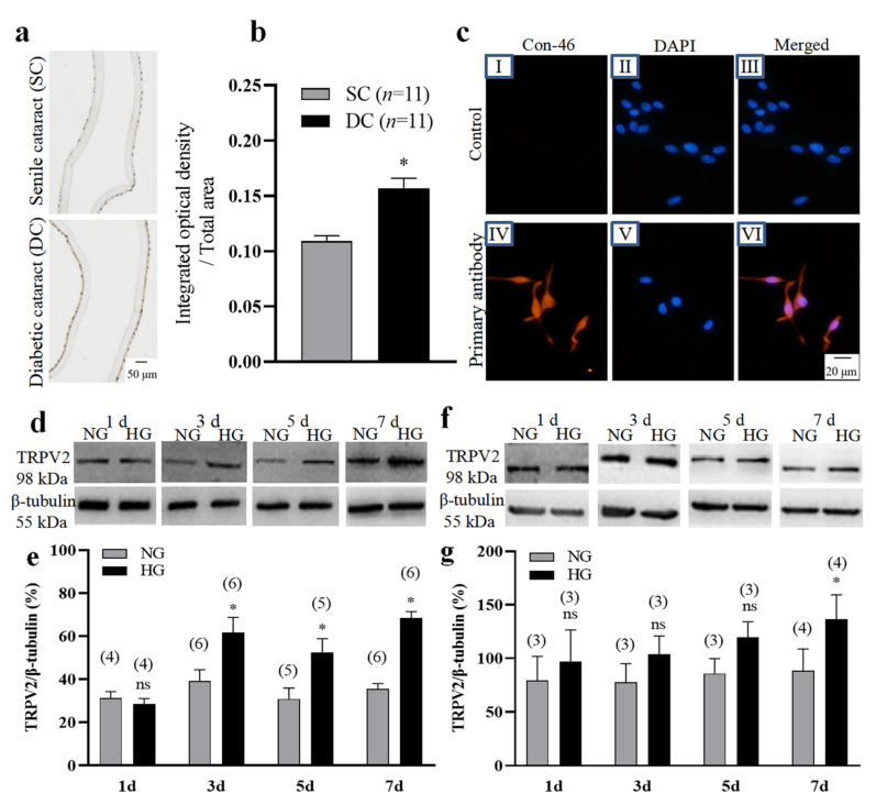
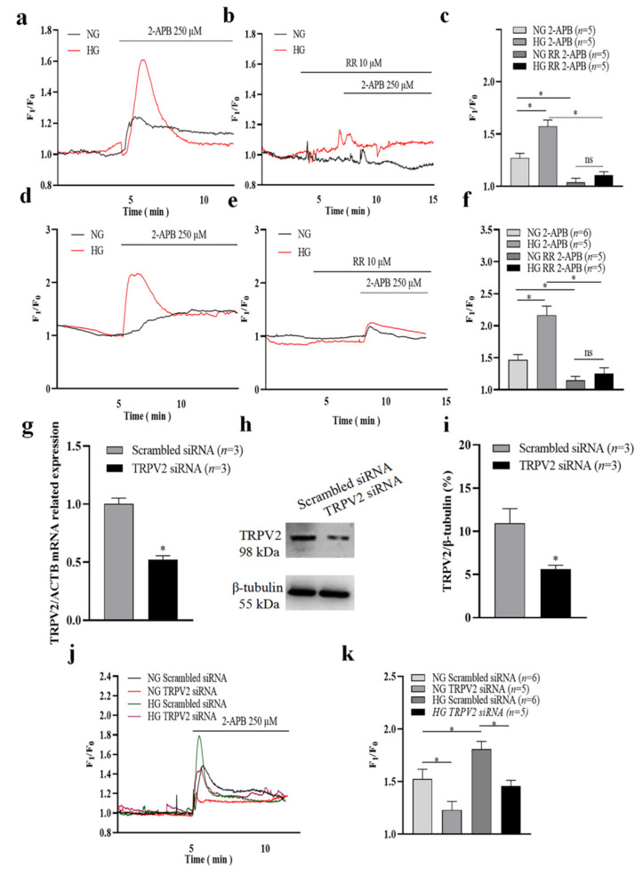
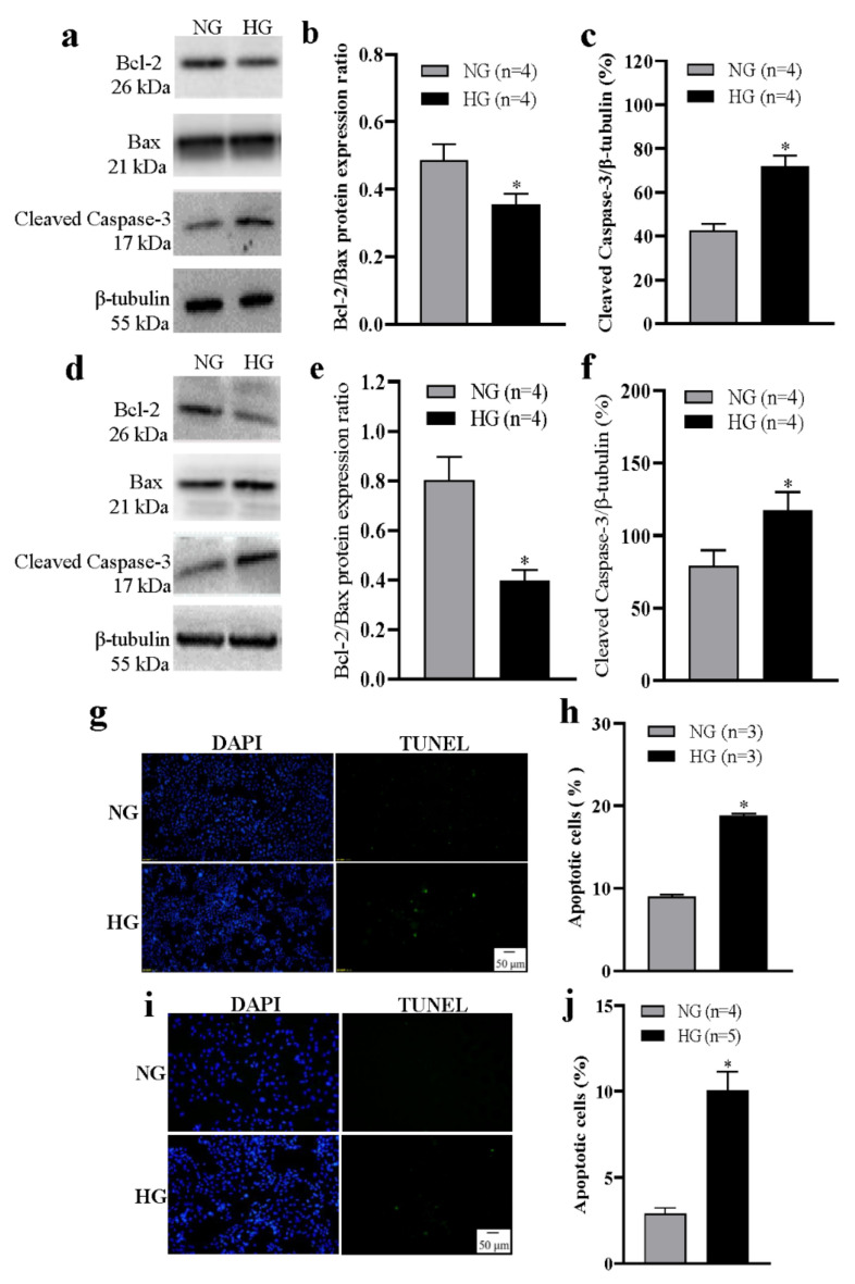
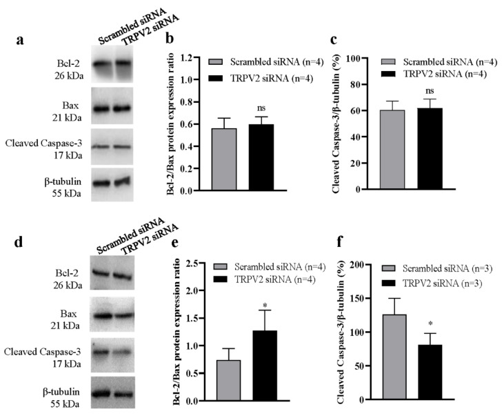
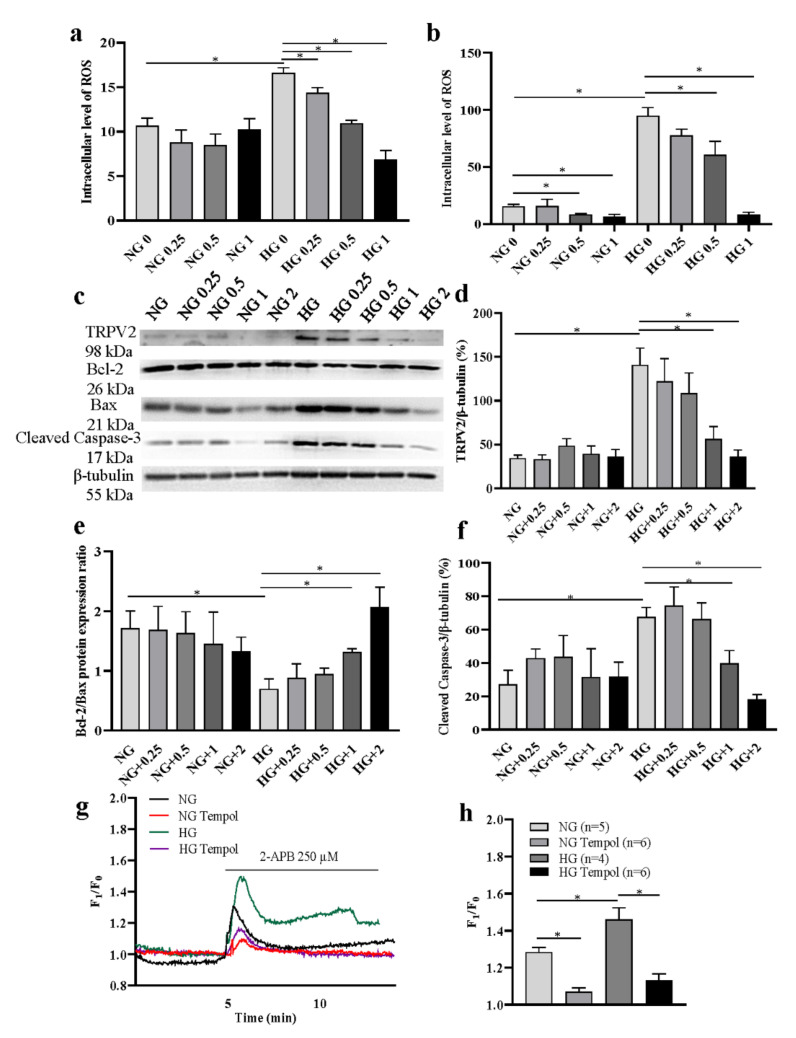
Cataracts are a serious complication of diabetes. In long-term hyperglycemia, intracellular Ca2+ concentration ([Ca2+]i) and reactive oxygen species (ROS) are increased. The apoptosis of lens epithelial cells plays a key role in the development of cataract. We investigated a potential role for transient receptor potential vanilloid 2 (TRPV2) in the development of diabetic cataracts. Immunohistochemical and Western blotting analyses showed that TRPV2 expression levels were significantly increased in the lens epithelial cells of patients with diabetic cataracts as compared with senile cataract, as well as in both a human lens epithelial cell line (HLEpiC) and primary rat lens epithelial cells (RLEpiCs) cultured under high-glucose conditions. The [Ca2+]i increase evoked by a TRPV2 channel agonist was significantly enhanced in both HLEpiCs and RLEpiCs cultured in high-glucose media. This enhancement was blocked by the TRPV2 nonspecific inhibitor ruthenium red and by TRPV2-specific small interfering (si)RNA transfection. Culturing HLEpiCs or RLEpiCs for seven days in high glucose significantly increased apoptosis, which was inhibited by TRPV2-specific siRNA transfection. In addition, ROS inhibitor significantly suppressed the ROS-induced increase of TRPV2-mediated Ca2+ signal and apoptosis under high-glucose conditions. These findings suggest a mechanism underlying high-glucose-induced apoptosis of lens epithelial cells, and offer a potential target for developing new therapeutic options for diabetes-related cataracts.

Keywords: TRPV2; apoptosis; diabetic cataract; human lens epithelial cell.


Conflict of interest statement

The authors declare no conflict of interest.

Figures

Figure 1
Figure 1
Changes in TRPV2 protein expression. (a,b) Representative images (a) and summary data (b) showing TRPV2 expression levels in human lens epithelial cells from patients having senile cataracts (SC) or diabetic cataracts (DC). In (b), data showing the ratio of integrated optical density/total area of TRPV2. (c) Immunofluorescence images showing the expression of connexin-46 (Con-46) protein in primary cultured lens epithelial cells from rat. No—primary antibody control: I–III Connexin-46 expression: IV-VI. Cell nuclei are stained blue by 4′,6-diamidino-2-phenylindole (DAPI). (dg) Representative Western blotting images (d,f) and summary data (e,g) showing the expression levels of TRPV2 in HLEpiCs (d,e) and primary rat lens epithelial cells (f,g) cultured in a normal-glucose (NG, 5.5 mM glucose and 20 mM mannitol) or high-glucose (HG, 25.6 mM glucose) media for 1, 3, 5, and 7 days. Numbers in parentheses over the bars in panels (e,g) represent the number of biological replicates. Values are shown as the mean ± SEM; n = 3–11. * p < 0.05 vs. SC or NG at 3, 5, and 7 days analyzed by two-tailed unpaired Student’s t test; ns, not significant.
Figure 2
Figure 2
Changes of TRPV2-mediated Ca2+ influx in a high-glucose environment. (af) Representative traces (a,b,d,e) and summary data (c,f) showing the changes in the intracellular Ca2+ concentration of HLEpiCs (ac) and primary rat lens epithelial cells (df) cultured in a normal-glucose (NG, 5.5 mM glucose and 20 mM mannitol) or high-glucose (HG, 25.6 mM glucose) medium for seven days. The cells were treated without or with ruthenium red (RR, 10 μM) and activated by 2-aminoethyldiphenyl borate (2-APB, 250 μM). (g) Summary data showing TRPV2 mRNA expression level of HLEpiCs transfected with TRPV2 siRNA or scrambled siRNA control for three days. (hi) Representative Western blotting images (h) and summary data (i) showing TRPV2 protein expression levels of HLEpiCs transfected with TRPV2 siRNA or scrambled siRNA control for three days. (jk) Representative traces (j) and summary data (k) of the changes in intracellular Ca2+ concentration of HLEpiCs transfected with TRPV2 siRNA or scrambled siRNA control and cultured in NG or HG media for seven days. The TRPV2 channel agonist 2-APB (250 μM) activated the channel to induce Ca2+ influx. Values are shown as the mean ± SEM; n = 3–6. * p < 0.05 analyzed by two-way analysis of variance followed by a Bonferroni test in panel c, f and k, and two-tailed unpaired Student’s t test in panel (g,i); ns, not significant.
Figure 3
Figure 3
High glucose–induced apoptosis of lens epithelial cells. (af) Representative Western blotting images (a,d) and summary data (b,c,e,f) showing the Bcl-2/Bax protein expression ratio (b,e) and the expression levels of active caspase-3 (c,f) in HLEpiCs (ac) and primary rat lens epithelial cells (df) cultured in a normal-glucose (NG, 5.5 mM glucose and 20 mM mannitol) or high-glucose (HG, 25.6 mM glucose) medium for seven days. (g,i) Representative images showing cell nuclei (blue, 4′,6-diamidino-2-phenylindole [DAPI]) and apoptotic HLEpiCs (g) and primary rat lens epithelial cells (i) (green, terminal deoxynucleotidyl transferase–mediated nick-end labeling) cultured in NG or HG media for seven days. (h,j) Summary data showing the percentage of apoptotic HLEpiCs (h) and primary rat lens epithelial cells (j). Values are shown as the mean ± SEM; n = 3–5. * p < 0.05 vs. NG analyzed by a two-tailed unpaired Student’s t test.
Figure 4
Figure 4
Role of TRPV2 in high glucose–induced apoptosis of lens epithelial cells. Representative Western blotting images (a,d) and summary data (b,c,e,f) showing Bcl-2/Bax protein expression ratios and the levels of active caspase-3 in HLEpiCs cultured in a normal-glucose ((ac), 5.5 mM glucose and 20 mM mannitol) or high-glucose ((e,f), 25.6 mM glucose) medium for seven days and transfected with TRPV2 siRNA or scrambled siRNA control. Values are shown as the mean ± SEM; n = 3–4. * p < 0.05 vs. scrambled siRNA analyzed by two-tailed unpaired Student’s t test; ns, not significant.
Figure 5
Figure 5
Effects of reactive oxygen species (ROS) on TRPV2 expression and function. (a,b) Summary data showing intracellular ROS level in HLEpiCs treated with different concentrations of an antioxidant Tempol (0, 0.25, 0.5, 1 mM) for three and seven days (n = 4). (cf) Representative Western blotting images (c) and summary data (df) showing the expression levels of TRPV2 (n = 4) (d), Bcl-2/Bax (n = 3) protein expression ratio (e) and active caspase-3 in HLEpiCs (f) cultured in a normal-glucose (NG, 5.5 mM glucose and 20 mM mannitol) or high-glucose (HG, 25.6 mM glucose) media treated with Tempol (0, 0.25, 0.5, 1, 2 mM). (g,h) Representative traces (g) and summary data (h) showing the changes in the intracellular Ca2+ concentration of HLEpiCs treated with Tempol (1 mM) cultured in a NG or HG medium for seven days. The cells were activated by 2-aminoethyldiphenyl borate (2-APB, 250 μM). Values are shown as the mean ± SEM; n = 3–6. * p < 0.05 analyzed by two-way analysis of variance, followed by a Bonferroni test.

References

    1. Ramsey I.S., Delling M., Clapham D.E. An introduction to TRP channels. Ann. Rev. Physiol. 2006;68:619–647. doi: 10.1146/annurev.physiol.68.040204.100431. - DOI - PubMed
    1. Benham C.D., Gunthorpe M.J., Davis J.B. TRPV channels as temperature sensors. Cell Calcium. 2003;33:479–487. doi: 10.1016/S0143-4160(03)00063-0. - DOI - PubMed
    1. Dietrich A., Chubanov V., Kalwa H., Rost B.R., Gudermann T. Cation channels of the transient receptor potential superfamily: Their role in physiological and pathophysiological processes of smooth muscle cells. Pharmacol. Ther. 2006;112:744–760. doi: 10.1016/j.pharmthera.2006.05.013. - DOI - PubMed
    1. Bravo-Sagua R., Parra V., López-Crisosto C., Díaz P., Quest A.F., Lavandero S. Calcium Transport and Signaling in Mitochondria. Compr. Physiol. 2017;7:623–634. doi: 10.1002/cphy.c160013. - DOI - PubMed
    1. Borowiec A.S., Bidaux G., Pigat N., Goffin V., Bernichtein S., Capiod T. Calcium channels, external calcium concentration and cell proliferation. Eur. J. Pharmacol. 2014;739:19–25. doi: 10.1016/j.ejphar.2013.10.072. - DOI - PubMed