. 2022 Apr 3;11(7):1210.
doi: 10.3390/cells11071210.

Equilibrium among Inflammatory Factors Determines Human MSC-Mediated Immunosuppressive Effect

Affiliations

Equilibrium among Inflammatory Factors Determines Human MSC-Mediated Immunosuppressive Effect

Yulia Suzdaltseva et al. Cells. .

Abstract

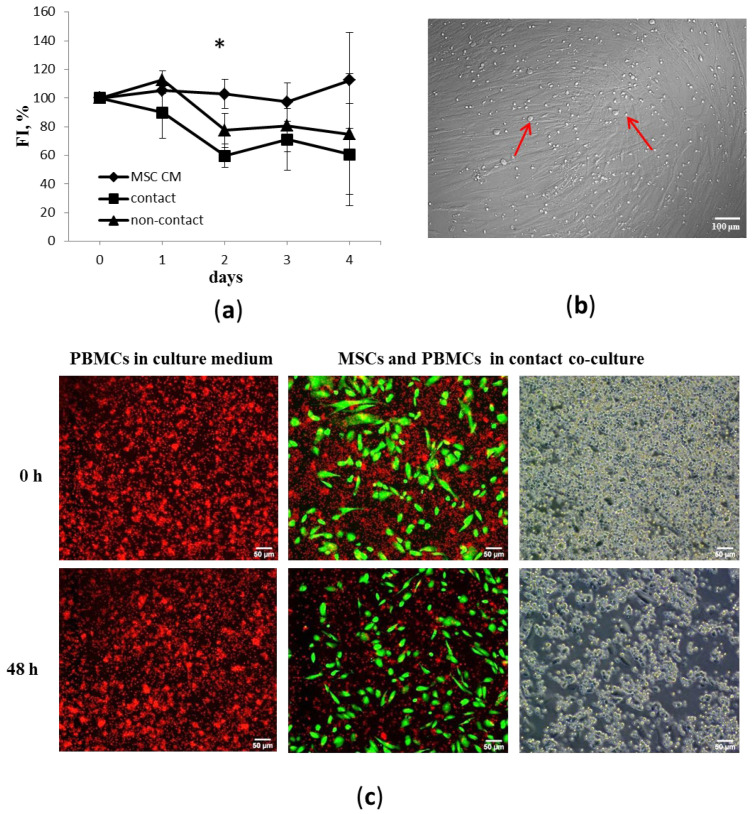
Mesenchymal stem cells (MSCs) are thought to be a promising therapeutic agent due to their multiple paracrine and immunomodulatory properties, providing protection from chronic inflammation and promoting tissue repair. MSCs can regulate the balance of pro-inflammatory and anti-inflammatory factors in inflamed tissues, creating a microenvironment necessary for successful healing; however, their interactions with immune cells are still poorly studied. We examined the temporal and spatial changes in gene regulation and the paracrine milieu accompanying the MSC-mediated immunosuppression effect in mixed cultures with activated peripheral blood mononuclear cells (PBMCs). Our data reveal that the peak of suppression of PBMC proliferation was achieved within 48 h following co-culture with MSCs and subsequently did not undergo a significant change. This effect was accompanied by an increase in COX-2 expression and an induction of IDO synthesis in MSCs. At this point, the expression of IL-1, IL-6, IL-8, IFN-γ, MCP-1, and G-CSF was upregulated in co-cultured cells. On the contrary, we observed a decrease in the concentrations of IL-10, IL-13, IL-5, and MIP-1b in co-culture supernatants compared to intact cultures of activated PBMCs. The regulation of IDO, IL-1, IL-6, and G-CSF production was accomplished with the involvement of direct cell-cell contact between MSCs and PBMCs. These findings provide new insights into the use of potential precondition inducers or their combinations to obtain functionally qualified MSCs for more effective treatment of inflammatory diseases.

Keywords: cytokines; immunomodulation; mesenchymal stem cells; priming.


Conflict of interest statement

The authors declare no conflict of interest.

Figures

Figure 1
Figure 1
Morphology, phenotype, and multilineage differentiation potential of adipose-derived MSCs. (a) Phase contrast image shows fibroblast-like morphology of MSCs from adhesion culture of second passage; (b) accumulation of mineralized matrix in MSCs, with alizarin red staining; (c) accumulation of adipose vacuoles in MSCs, with Oil Red O and hematoxylin staining; (d) toluidine blue staining of mucopolysaccharide extracellular matrix in cryosections from MSC micromass pellets cultured in chondrogenic media; and (e) flow fluorometry of MSCs stained with antibodies to surface markers CD90, CD73, CD105 CD34, CD45, and CD14 conjugated with Alexa Fluor 647, APC, and Per-CP-Cy5.5, with green indicating isotype control and red indicating specific surface markers.
Figure 2
Figure 2
Effects of MSCs on PBMC proliferation. (a) Dynamics of PHA-induced proliferation of PBMCs incubated with MSC conditioned medium (MSC CM) or adherent MSCs in contact or non-contact co-cultures at a 25:1 ratio, respectively. Five unrelated donors per cell type were used. Average values and standard errors from five independent fluorescence intensity measurements using CyQUANT® NF (FI) are shown. Data are normalized to the level of proliferation (100%) of PBMCs cultured in RPMI-1640 medium. Asterisk (*) indicates the reliability of differences at p < 0.05. (b) Red arrows indicate immune cells that formed close contacts with MSCs. Representative images of MSC and PBMC co-cultures after removing supernatant with nonadherent immune cells, phase contrast. (c) Representative images of live cells showing suppressive effect of MSCs (green) on PBMC proliferation (red) (CellTrace ™ calcein green and red-orange AM staining).
Figure 3
Figure 3
COX-2, IDO, and iNOS expression in MSCs in contact and non-contact (tw) co-culture with PHA-activated PBMCs. (a,b) Relative changes in COX-2 and IDO expression; mean values and their errors from 3 independent experiments are presented. MSCs were isolated from 3 unrelated donors. * reliability of differences at p < 0.05. (c) Representative electropherograms of PCR products.
Figure 4
Figure 4
Evaluation of human cytokine concentration in conditioned medium of MSCs and PHA-activated PBMCs, determined via multiplexed fluorescent bead-based immunoassay detection. Three unrelated donors per cell type were used. Data are shown as mean ± std; t-test, * reliability of differences at p < 0.05.
Figure 5
Figure 5
IL-1 concentration in conditioned media of separate MSC and activated PBMC cultures or contact and noncontact (tw) co-cultures. Data are shown as mean ± std; t-test, * reliability of differences at p < 0.05. Three unrelated donors per cell type were used.
Figure 6
Figure 6
Suppression of PBMC proliferation in the presence of MSCs accompanied by enhanced production of (a) IL-1, (b) IFN-γ, (c) IL-6, and (d) G-CSF. Data are shown as mean ± std; n = 5, t-test, * reliability of differences at p < 0.05. Contact refers to contact MSC–PBMC co-culture, tw refers to non-contact co-culture. Five unrelated donors per cell type were used.
Figure 7
Figure 7
Production of (a) IL-10, (b) IL-13, (c) IL-5, and (d) MIP-1b by activated PBMCs is inhibited in contact and non-contact (tw) co-cultures with MSCs. Data are shown as mean ± std; n = 5, t-test, * reliability of differences at p < 0.05. Contact refers to contact MSC–PBMC co-culture, tw refers to non-contact co-culture. Five unrelated donors per cell type were used.

References

    1. Pittenger M.F., Discher D.E., Péault B.M., Phinney D.G., Hare J.M., Caplan A.I. Mesenchymal stem cell perspective: Cell biology to clinical progress. NPJ Regen. Med. 2019;4:22. doi: 10.1038/s41536-019-0083-6. - DOI - PMC - PubMed
    1. Suzdaltseva Y., Zhidkih S., Kiselev S.L., Stupin V. Locally Delivered Umbilical Cord Mesenchymal Stromal Cells Reduce Chronic Inflammation in Long-Term Nonhealing Wounds: A Randomized Study. Stem Cells Int. 2020;2020:5308609. doi: 10.1155/2020/5308609. - DOI - PMC - PubMed
    1. Shi Y., Wang Y., Li Q., Liu K., Hou J., Shao C., Wang Y. Immunoregulatory mechanisms of mesenchymal stem and stromal cells in inflammatory diseases. Nat. Rev. Nephrol. 2018;14:493–507. doi: 10.1038/s41581-018-0023-5. - DOI - PubMed
    1. Zhou Y., Yamamoto Y., Xiao Z., Ochiya T. The Immunomodulatory Functions of Mesenchymal Stromal/Stem Cells Mediated via Paracrine Activity. J. Clin. Med. 2019;8:1025. doi: 10.3390/jcm8071025. - DOI - PMC - PubMed
    1. Waterman R.S., Tomchuck S.L., Henkle S.L., Betancourt A.M. A New Mesenchymal Stem Cell (MSC) Paradigm: Polarization into a Pro-Inflammatory MSC1 or an Immunosuppressive MSC2 Phenotype. PLoS ONE. 2010;5:e10088. doi: 10.1371/journal.pone.0010088. - DOI - PMC - PubMed

Publication types

MeSH terms