Review
. 2022 Apr 6;11(7):1239.
doi: 10.3390/cells11071239.

The Influence of Gut Dysbiosis in the Pathogenesis and Management of Ischemic Stroke

Affiliations
Review

The Influence of Gut Dysbiosis in the Pathogenesis and Management of Ischemic Stroke

Saravana Babu Chidambaram et al. Cells. .

Abstract

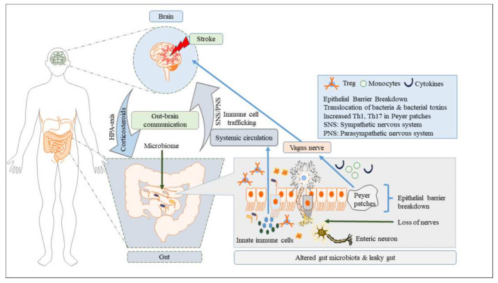
Recent research on the gut microbiome has revealed the influence of gut microbiota (GM) on ischemic stroke pathogenesis and treatment outcomes. Alterations in the diversity, abundance, and functions of the gut microbiome, termed gut dysbiosis, results in dysregulated gut-brain signaling, which induces intestinal barrier changes, endotoxemia, systemic inflammation, and infection, affecting post-stroke outcomes. Gut-brain interactions are bidirectional, and the signals from the gut to the brain are mediated by microbially derived metabolites, such as trimethylamine N-oxide (TMAO) and short-chain fatty acids (SCFAs); bacterial components, such as lipopolysaccharide (LPS); immune cells, such as T helper cells; and bacterial translocation via hormonal, immune, and neural pathways. Ischemic stroke affects gut microbial composition via neural and hypothalamic-pituitary-adrenal (HPA) pathways, which can contribute to post-stroke outcomes. Experimental and clinical studies have demonstrated that the restoration of the gut microbiome usually improves stroke treatment outcomes by regulating metabolic, immune, and inflammatory responses via the gut-brain axis (GBA). Therefore, restoring healthy microbial ecology in the gut may be a key therapeutic target for the effective management and treatment of ischemic stroke.

Keywords: cerebral stroke; gut dysbiosis; gut immune cells; gut leakiness; gut microbiota; gut-derived metabolites; gut–brain axis.


Conflict of interest statement

The authors declare that they have no conflict of interest.

Figures

Figure 1
Figure 1
Effects of stroke on the gut–brain axis. Following cerebral stroke, gut dysbiosis causes loss of enteric nerves, increased intestinal-barrier permeability, reduced mucus production, loss of goblet cells, thinning of the mucus barrier, and increased sympathetic activity in the intestinal wall, all of which contributes to intestinal inflammation and an exaggerated immune response. These events in turn disrupt intestinal and systemic immune homeostasis, resulting in poor stroke treatment prognosis.
Figure 2
Figure 2
Effect of gut dysbiosis following cerebral ischemic/reperfusion injury on brain structure and function. The eight brain-specific regions, such as the orbitofrontal cortex (OC), somatosensory cortex (SC), cingulate cortex (CC), hippocampus (H), motor cortex (MC), thalamus (T), auditory cortex (AC), and visual cortex (VC), in mice subjected to cerebral ischemic/reperfusion injury induced by occlusion of bilateral carotid common carotid arteries (BCCAO group) or sham-operation (control group). (A) The functional connectivity between specific regions of interest of both control and BCCAO groups is shown in the virtual graphics. (B) Mean functional connectivity strength in brain network was measured using two-way repeated-measures ANOVA with Tukey multiple comparisons as post hoc test. (C) The mean functional connectivity matrices show the strength of functional connectivity between pairs of brain regions in the normal and stroke groups. (D) Correlation analyses in the regions (8) of interest in animal brain; the colour scale indicates the functional connectivity strength. * p < 0.05. The figure is reused as per journal copyright permission [145].
Figure 3
Figure 3
(A,B) Metagenomic analysis of mice in BCCAO and control groups showed significant changes in gut microbial composition. (C) Firmicutes/Bacteroidetes ratio was reduced in BCCAO vs. control groups on day 29 after FMT. (D) The effects of baicalin treatment on the gut microbial populations at the phylum level showed that the plot of principal component 1 against principal component 2 formed a distinct cluster. Phylum clustering in BCCAO group was different from that in the control group. *** p < 0.01. The figure is reused as per journal copyright permission [145].
Figure 4
Figure 4
The relative taxa abundance between cerebral ischemic stroke patients (n = 30) and healthy controls (n = 30) belong to three phyla: Bacteroidetes, Firmicutes, and Proteobacteria (a) and to 25 genera that comprise up to 80% of the total microbiota, such as Bacteroides, Prevotella, Faecalibacterium, Escherichia/Shigella, Phascolarctobacterium, and Roseburia (b). The figure is reused as per journal copyright permission [113].
Figure 5
Figure 5
Heat map of Spearman correlation coefficient analysis, including the National Institutes of Health Stroke Scale (NIHSS) after 7 and 30 days and modified Rankin scale (mRS), was used to explore the relationship between the gut microbiota and severity or outcome of cerebral ischemic stroke patients. * p < 0.05, ** p < 0.01. The figure is reused as per journal copyright permission [113].

References

    1. Parr E., Ferdinand P., Roffe C. Management of Acute Stroke in the Older Person. Geriatrics. 2017;2:27. doi: 10.3390/geriatrics2030027. - DOI - PMC - PubMed
    1. Yamashiro K., Tanaka R., Urabe T., Ueno Y., Yamashiro Y., Nomoto K., Takahashi T., Tsuji H., Asahara T., Hattori N. Gut Dysbiosis Is Associated with Metabolism and Systemic Inflammation in Patients with Ischemic Stroke. PLoS ONE. 2017;12:e0171521. doi: 10.1371/journal.pone.0171521. - DOI - PMC - PubMed
    1. Benjamin E.J., Virani S.S., Callaway C.W., Chamberlain A.M., Chang A.R., Cheng S., Chiuve S.E., Cushman M., Delling F.N., Deo R., et al. Heart Disease and Stroke Statistics-2018 Update: A Report from the American Heart Association. Circulation. 2018;137:e67–e492. doi: 10.1161/CIR.0000000000000558. - DOI - PubMed
    1. Hossmann K.-A. Pathophysiology and Therapy of Experimental Stroke. Cell Mol. Neurobiol. 2006;26:1057–1083. doi: 10.1007/s10571-006-9008-1. - DOI - PMC - PubMed
    1. Li X., Li X., Lin H., Fu X., Lin W., Li M., Zeng X., Gao Q. Metabolic Syndrome and Stroke: A Meta-Analysis of Prospective Cohort Studies. J. Clin. Neurosci. 2017;40:34–38. doi: 10.1016/j.jocn.2017.01.018. - DOI - PubMed

Publication types

Substances