

A Comparison of the Genotoxic Effects of Gold Nanoparticles Functionalized with Seven Different Ligands in Cultured Human Hepatocellular Carcinoma Cells
- PMID: 35407243
- PMCID: PMC9000686
- DOI: 10.3390/nano12071126
A Comparison of the Genotoxic Effects of Gold Nanoparticles Functionalized with Seven Different Ligands in Cultured Human Hepatocellular Carcinoma Cells
Abstract
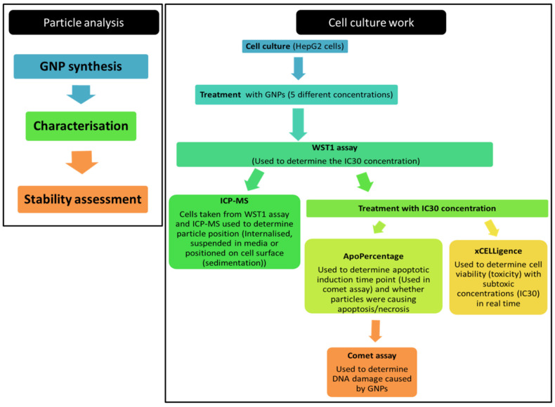
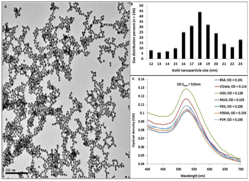
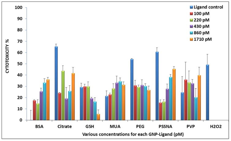
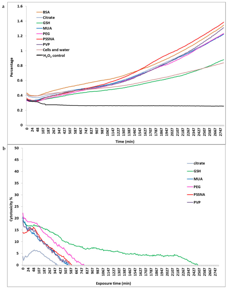
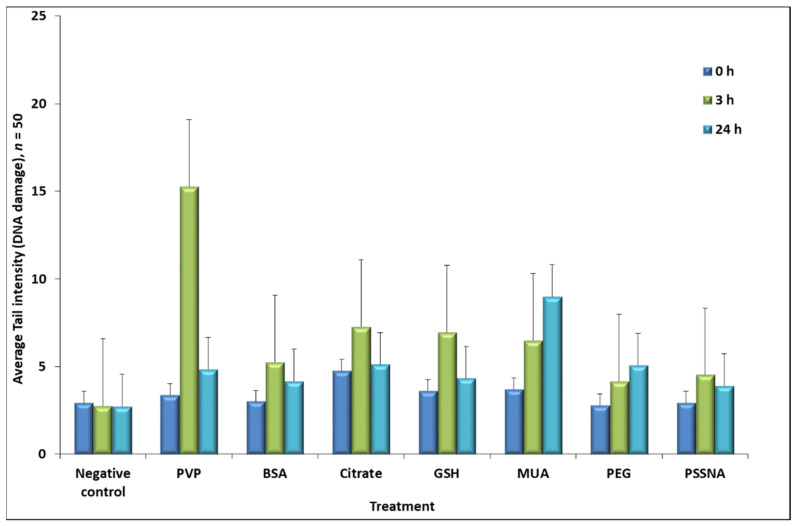
Gold nanoparticles (GNPs) have shown great potential in diagnostic and therapeutic applications in diseases, such as cancer. Despite GNP versatility, there is conflicting data regarding the toxicity of their overall functionalization chemistry for improved biocompatibility. This study aimed to determine the possible genotoxic effects of functionalized GNPs in Human hepatocellular carcinoma (HepG2) cells. GNPs were synthesized and biofunctionalized with seven common molecules used for biomedical applications. These ligands were bovine serum albumin (BSA), poly(sodium 4-styrene sulfonate) (PSSNA), trisodium citrate (citrate), mercaptoundecanoic acid (MUA), glutathione (GSH), polyvinylpyrrolidone (PVP), and polyethylene glycol (PEG). Before in vitro genotoxicity assessment, inductively coupled plasma mass spectrometry was used to determine GNP cellular internalization quantitatively, followed by cell-based assays; WST-1 to find IC 30 and ApoPercentage for apoptotic induction time-points. The effect of the GNPs on cell growth in real-time was determined by using xCELLigence, followed by a comet assay for genotoxicity determination. The HepG2 cells experienced genotoxicity for all GNP ligands; however, they were able to initiate repair mechanisms and recover DNA damage, except for two functionalization chemistries.
Keywords: HepG2; biofunctionalization; comet assay; cytotoxicity; genotoxicity; gold nanoparticles.
Conflict of interest statement
The authors declare no conflict of interest.
Figures
References
-
- Solano-Umaña V., Vega Baudrit J. The New Field of the Nanomedicine. Int. J. Appl. Sci. Technol. 2015;5:79.
-
- Panyala R.N., Pena-Mendez M.E., Havel J. Gold and nano-gold in medicine: Overview, toxicology and perspectives. J. Appl. Biomed. 2009;7:75–91. doi: 10.32725/jab.2009.008. - DOI
-
- Rosarin F., Mirunalini S. Nobel metallic nanoparticles with novel biomedical properties. J. Bioanal. Biomed. 2011;3:85–91. doi: 10.4172/1948-593X.1000049. - DOI
LinkOut - more resources
Full Text Sources
