. 2022 Apr 3;11(7):2011.
doi: 10.3390/jcm11072011.

Standardized Classification of Cerebral Vasospasm after Subarachnoid Hemorrhage by Digital Subtraction Angiography

Affiliations

Standardized Classification of Cerebral Vasospasm after Subarachnoid Hemorrhage by Digital Subtraction Angiography

Helena Merkel et al. J Clin Med. .

Abstract

Background: During the last decade, cerebral vasospasm after aneurysmal subarachnoid hemorrhage (SAH) was a current research focus without a standardized classification in digital subtraction angiography (DSA). This study was performed to investigate a device-independent visual cerebral vasospasm classification for endovascular treatment.

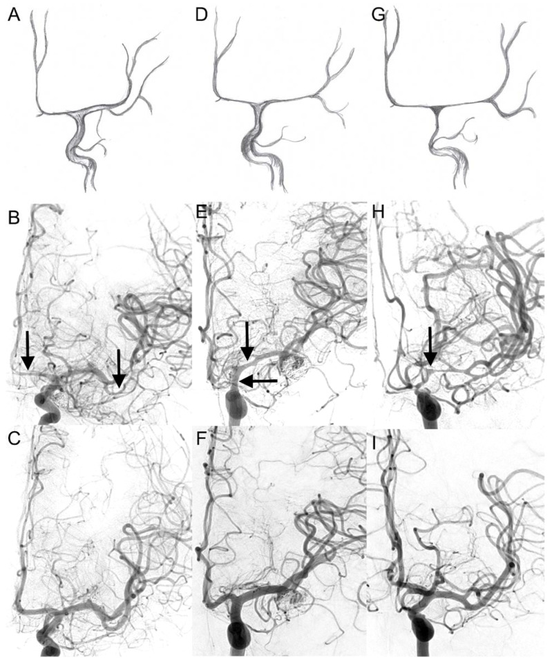
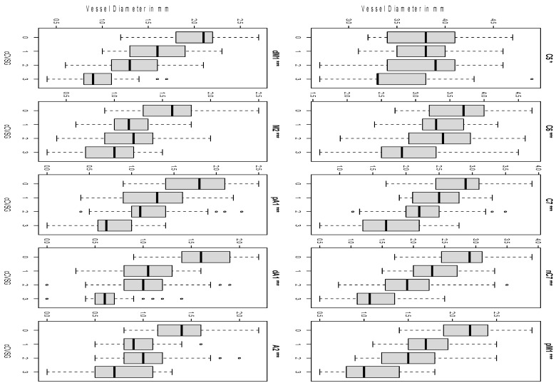
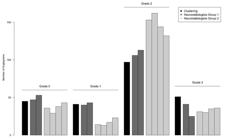
Methods: The analyses are DSA based rather than multimodal. Ten defined points of intracranial arteries were measured in 45 patients suffering from cerebral vasospasm after SAH at three time points (hospitalization, before spasmolysis, control after six months). Mathematical clustering of vessel diameters was performed to generate four objective grades for comparison. Six interventional neuroradiologists in two groups scored 237 DSAs after a new visual classification (grade 0-3) developed on a segmental pattern of vessel contraction. For the second group, a threshold-based criterion was amended.

Results: The raters had a reproducibility of 68.4% in the first group and 75.2% in the second group. The complementary threshold-based criterion increased the reproducibility by about 6.8%, while the rating deviated more from the mathematical clustering in all grades.

Conclusions: The proposed visual classification scheme of cerebral vasospasm is suitable as a standard grading procedure for endovascular treatment. There is no advantage of a threshold-based criterion that compensates for the effort involved. Automated vessel analysis is superior to compare inter-group results in research settings.

Keywords: cerebral vasospasm; classification; subarachnoid hemorrhage; vessel diameter.


Conflict of interest statement

The authors declare no conflict of interest.

Figures

Figure 1
Figure 1
Visual cerebral vasospasm classification in digital subtraction angiography. The upper two lines display cerebral vasospasm grades (CVSG) as drawings with the corresponding angiograms. In the bottom line, the corresponding control DSA after six months is shown: (A,B) CVSG 1—Narrowing of the A2, A1, and M2 segments (arrows) with postspastic enlargement of distal M2 branches. (D,E) CVSG 2—The spasm affects the proximal M1 segment and the intradural carotid artery (arrows). (G,H) CVSG 3—The intradural carotid artery, the proximal middle cerebral artery, and the anterior cerebral artery show high-grade narrowing with a fading appearance like a ghost (arrow). (C,F,I)—control DSA after 6 months (CVSG 0).
Figure 2
Figure 2
Clustering of vessel diameters. The distribution of vessel diameters according to cCVSG 0–3 after mathematical clustering is shown. All vessel segments had a significant intergrade difference (* C5 p = 0.013, *** the remaining p < 0.001). Discordant values are shown as small circles C5 clinoid segment of the internal carotid artery, C6 ophthalmic segment of the internal carotid artery, C7 terminal segment of the internal carotid artery, nC7 narrowest point of C7, pM1 proximal horizontal segment of the middle cerebral artery, dM1 distal horizontal segment of the middle cerebral artery, M2 insular segment of the middle cerebral artery, pA1 proximal pre-communication segment of the anterior cerebral artery, dA1 distal pre-communication segment of the anterior cerebral artery, A2 post-communicating segment of the anterior cerebral artery. The small circles show the discordant values.
Figure 3
Figure 3
Reproducibility of visual classification of cerebral vasospasm. The mathematical clustering for objective comparison is depicted in black first, followed by the subjective grading of each rater labeled according to his profession. The first group in dark gray (two neuroradiologists) applied the primary visual cerebral vasospasm classification. The second group in light gray (four neuroradiologists) applied the modified classification supplemented with the pM1 values. Neuroradiologists of the second group deviate more from the clustering.

References

    1. Francoeur C.L., Mayer S.A. Management of delayed cerebral ischemia after subarachnoid hemorrhage. Crit. Care. 2016;20:277. doi: 10.1186/s13054-016-1447-6. - DOI - PMC - PubMed
    1. Yao Z., Hu X., You C. Endovascular therapy for vasospasm secondary to subarachnoid hemorrhage: A meta-analysis and systematic review. Clin. Neurol. Neurosurg. 2017;163:9–14. doi: 10.1016/j.clineuro.2017.09.016. - DOI - PubMed
    1. Connolly E.S., Jr., Rabinstein A.A., Carhuapoma J.R., Derdeyn C.P., Dion J., Higashida R.T., Hoh B.L., Kirkness C.J., Naidech A.M., Ogilvy C.S., et al. Guidelines for the management of aneurysmal subarachnoid hemorrhage: A guideline for healthcare professionals from the American Heart Association/american Stroke Association. Stroke. 2012;43:1711–1737. doi: 10.1161/STR.0b013e3182587839. - DOI - PubMed
    1. Janjua N., Mayer S.A. Cerebral vasospasm after subarachnoid hemorrhage. Curr. Opin. Crit. Care. 2003;9:113–119. doi: 10.1097/00075198-200304000-00006. - DOI - PubMed
    1. Samagh N., Bhagat H., Jangra K. Monitoring cerebral vasospasm: How much can we rely on transcranial Doppler. J. Anaesthesiol. Clin. Pharmacol. 2019;35:12–18. doi: 10.4103/joacp.JOACP_192_17. - DOI - PMC - PubMed

LinkOut - more resources