. 2022 Apr 5;15(7):2671.
doi: 10.3390/ma15072671.

Design of New Concept of Knitted Hernia Implant

Affiliations

Design of New Concept of Knitted Hernia Implant

Bogusława Żywicka et al. Materials (Basel). .

Abstract

A knitted implant, unilaterally modified with plasma-assisted chemical-vapor deposition (PACVD), and with a nano-layer of fluorine derivative supplementation, for reducing the risk of complications related to adhesions, and the formation of a thick postoperative scar was prepared. The biological evaluation of designed or modified medical devices is the main aspect of preclinical research. If such studies use a medical device with prolonged contact with connective tissue (more than 30 days), biocompatibility studies require a safety assessment in terms of toxicity in vitro and in vivo, allergenicity, irritation, and cancerogenicity, reproductive and developmental toxicity. The ultimate aspect of biological evaluation is biofunctionality, and evaluation of the local tissue response after implantation, resulting in the determination of all aspects of local biocompatibility with the implemented synthetic material. The implantation of PACVD-modified materials in muscle allows us to estimate the local irritation effect on the connective tissue, determining the risk of scar formation, whereas implantation of the above-mentioned knitted fabric into the abdominal wall, assists with evaluating the risk of fistula formation-the main post-surgical complications. The research aimed to evaluate the local reaction of the soft tissues after the implantation of the knitted implants modified with PACVD of the fluoropolymer in the nanostuctural form. The local effect that occurred during the implantation of the designed implants was quantitatively and qualitatively evaluated when PACVD unmodified (reference), and modified medical devices were implanted in the abdominal cavity (intra-abdominal position) for 12 or into the muscles for 56 weeks. The comparative semi-quantitative histological assessment included the severity of inflammatory cells (multinucleated cells, lymphocytes, plasma cells, macrophages, giant cells) and the tissue response (necrosis, neovascularization, fibrosis, and fat infiltration) on a five-point scale. The knitted implants modified by PACVD did not indicate cumulative tissue response when they were implanted in the muscle and intra-abdominally with direct contact with the viscera. They reduced local tissue reaction (score -2.71 after 56 weeks of the implantation) and internal organ adhesion (irritation score -2.01 and adhesion susceptibility -0.3 after 12 weeks of the implantation) compared with the reference (unmodified by PACVD) knitted implant, which had an identical structure and was made of the same source.

Keywords: PACVD; hernia knitted implants; irritation; post-implantation effect; viscera adhesion prevention.


Conflict of interest statement

The authors declare no conflict of interest.

Figures

Figure 1
Figure 1
Optical microphotograph (a) and SEM microphotograph (mag, 800×) of the designed PACVD-modified prototype of the hernia implant (b) used in the study. The idea of PACVD-modification of the designed knitted implants: 1—the flat surface of the implant subjected to PACVD-modification; 2—surface not subjected to the modification with unfolding in the form of protruding loops (c) [35].
Figure 2
Figure 2
The knitted implants used to estimate the local effect after implantation: (a) materials for the test used; (b) macroscopic view of the surface with loops; (c) macroscopic view of the opposite surface without loops and PACVD-modified with the presence of the fluoropolymer.
Figure 3
Figure 3
Intrasurgical photography: implantation of samples into pockets in the dorsal muscles of an NZW rabbit. The evaluated samples are localized on the left (a), and the reference on the right side of the spine (b).
Figure 4
Figure 4
Intrasurgical photography: implantation of the tested implant into the abdominal wall (intraperitoneal position): (a)—the operative filed view; (b)—location of the implant.
Figure 5
Figure 5
Macroscopic view of: (a)—the tested PACVD-modified sample and (b)—after 12 weeks of intramuscular implantation (implants are marked by arrows.
Figure 6
Figure 6
A microscopic view of the PACVD-modified sample (a) and the reference (b) 1 week after the intramuscular implantation. The monofilaments of knitted fabrics (i) surrounded by a loose connective tissue (lct) are separated from the muscle tissue (mt). A rich-in-cell, loose connective tissue (rlct) in the immediate vicinity of the monofilaments of the knitted structure is visible. Magnification 100×, Stain. HE and VG; Magnification 400×, Stain. HE.
Figure 7
Figure 7
A microscopic view of the PACVD modified sample (a) and the reference (b) 3 weeks after the intramuscular implantation. In the muscles (mt), the implant monofilaments are surrounded by connective tissue (fct) with numerous blood vessels (bv). A loose connective tissue with single giant cells in the immediate vicinity of the knitted structure is visible. Magnification 100×, Stain. VG; Magnification 400×, Stain. HE.
Figure 8
Figure 8
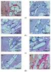
A microscopic view of the PACVD-modified sample (left side) and the reference (right side) after 9 (a), 12 (b), 26 (c), or 56 weeks (d) after the intramuscular implantation. In the muscle tissue (mt), the monofilaments of knitted implants (i) are surrounded by fibrous connective tissue (fct) with blood vessels (bv) and bands of fatty infiltrate (ft). Loose connective tissue (lct) with single polynuclear macrophages (gc) in the immediate vicinity of the implant structures (i) is visible. Magnification 100×, Stain. HE and VG.
Figure 9
Figure 9
A microscopic view of the PACVD-modified implant (a) and reference (b) 2 weeks after the implantation in the abdominal wall of a NZW rabbit. Monofilaments of knitted structure (i) are surrounded by loose connective tissue; image from the muscle tissue (mt) side (on the left) and from the peritoneal cavity (pc) (in the middle). In the immediate vicinity of the fibers, polynuclear macrophages (gc) are formed (on the right). Magnification, 100× and 400×, Stain. HE.
Figure 9
Figure 9
A microscopic view of the PACVD-modified implant (a) and reference (b) 2 weeks after the implantation in the abdominal wall of a NZW rabbit. Monofilaments of knitted structure (i) are surrounded by loose connective tissue; image from the muscle tissue (mt) side (on the left) and from the peritoneal cavity (pc) (in the middle). In the immediate vicinity of the fibers, polynuclear macrophages (gc) are formed (on the right). Magnification, 100× and 400×, Stain. HE.
Figure 10
Figure 10
A microscopic view of the PACVD-modified implant (a) and reference (b) 4 weeks after the implantation in the abdominal wall of a NZW rabbit. The monofilaments of knitted structures (i) are surrounded by fibrous connective tissue (fct) from the side of the muscles (on the left) of the abdominal wall and peritoneal cavity (pc) (in the middle). On the right side, the loose connective tissue (lct) with polynuclear macrophages (gc) appears in the immediate vicinity of the monofilament. Magnification, 100×, Stain. VG and HE. Magnification 400×, Stain. HE.
Figure 11
Figure 11
A microscopic view of the PACVD-modified implant (a) and reference (b) 12 weeks after the implantation in the abdominal wall of a NZW rabbit. Monofilaments of the knitted structure (i) are surrounded by connective tissue, which was fibrous (fct) from the muscles (mt) side and loose (lct) in the vicinity of the thread. On the right side, the adjacent intestinal wall (iw) is visible. Magnification, 100×, Stain. VG and HE.

References

    1. Otero J., Huber A.T., Heniford B.T. Laparoscopic Hernia Repair. Adv. Surg. 2019;53:1–19. doi: 10.1016/j.yasu.2019.04.001. - DOI - PubMed
    1. Corthals S., van Cleven S., Uyttebroek O., de Carvalho A., Vanlander A., Berrevoet F. Quality of Life After Open Versus Laparoscopic Preperitoneal Mesh Repair for Unilateral Inguinal Hernias. Asian J. Surg. 2021;44:1266–1273. doi: 10.1016/j.asjsur.2021.03.014. - DOI - PubMed
    1. Yu S., Ma P. Mechanical Properties of Warp-Knitted Hernia Repair Mesh with Various Boundary Conditions. J. Mech. Behav. Biomed. Mater. 2021;114:104192. doi: 10.1016/j.jmbbm.2020.104192. - DOI - PubMed
    1. Turlakiewicz K., Puchalski M., Krucinska I., Sujka W. The Role of Mesh Implants in Surgical Treatment of Parastomal Hernia. Materials. 2021;14:1062. doi: 10.3390/ma14051062. - DOI - PMC - PubMed
    1. Bendavid R., Abrahamson J., Arregui M.E., Flament J.B., Phillips E.H. Abdominal Wall Hernias: Principles And Management. Springer; Cham, Switzerland: 2018. pp. 39–192.

LinkOut - more resources