. 2022 Mar 28;27(7):2176.
doi: 10.3390/molecules27072176.

5-Bromo-3,4-dihydroxybenzaldehyde Promotes Hair Growth through Activation of Wnt/β-Catenin and Autophagy Pathways and Inhibition of TGF-β Pathways in Dermal Papilla Cells

Affiliations

5-Bromo-3,4-dihydroxybenzaldehyde Promotes Hair Growth through Activation of Wnt/β-Catenin and Autophagy Pathways and Inhibition of TGF-β Pathways in Dermal Papilla Cells

Jung-Il Kang et al. Molecules. .

Abstract

Various studies addressing the increasing problem of hair loss, using natural products with few side effects, have been conducted. 5-bromo-3,4-dihydroxybenzaldehyde (BDB) exhibited anti-inflammatory effects in mouse models of atopic dermatitis and inhibited UVB-induced oxidative stress in keratinocytes. Here, we investigated its stimulating effect and the underlying mechanism of action on hair growth using rat vibrissa follicles and dermal papilla cells (DPCs), required for the regulation of hair cycle and length. BDB increased the length of hair fibers in rat vibrissa follicles and the proliferation of DPCs, along with causing changes in the levels of cell cycle-related proteins. We investigated whether BDB could trigger anagen-activating signaling pathways, such as the Wnt/β-catenin pathway and autophagy in DPCs. BDB induces activation of the Wnt/β-catenin pathway through the phosphorylation of GSG3β and β-catenin. BDB increased the levels of autophagic vacuoles and autophagy regulatory proteins Atg7, Atg5, Atg16L, and LC3B. We also investigated whether BDB inhibits the TGF-β pathway, which promotes transition to the catagen phase. BDB inhibited the phosphorylation of Smad2 induced by TGF-β1. Thus, BDB can promote hair growth by modulating anagen signaling by activating Wnt/β-catenin and autophagy pathways and inhibiting the TGF-β pathway in DPCs.

Keywords: 5-bromo-3,4-dihydroxybenzaldehyde; TGF-β; Wnt/β-catenin; autophagy; dermal papilla cells; hair growth; proliferation.


Conflict of interest statement

The authors declare no conflict of interest.

Figures

Figure 1
Figure 1
BDB increases the length of hair fibers on the cultured hair follicles ex vivo. Rat vibrissa follicles were stimulated using BDB (0.01, 0.1, and 1 μM) or minoxidil (10 μM) for 21 days: (a) Structure of BDB. (b) Photograph of rat vibrissa follicles cultured at 0 and 21 days. (c) Changes in length of hair fibers on the vibrissa follicles treated with BDB or minoxidil for 21 days. The bar graph shows the growth percentage compared to the average length in the control group on day 21. Each dot indicates independent length of hair fibers on the vibrissa follicle (%). Data are presented as mean ± SD. * p < 0.05 compared with the control. The red word (Korean word) in the (b) means length.
Figure 1
Figure 1
BDB increases the length of hair fibers on the cultured hair follicles ex vivo. Rat vibrissa follicles were stimulated using BDB (0.01, 0.1, and 1 μM) or minoxidil (10 μM) for 21 days: (a) Structure of BDB. (b) Photograph of rat vibrissa follicles cultured at 0 and 21 days. (c) Changes in length of hair fibers on the vibrissa follicles treated with BDB or minoxidil for 21 days. The bar graph shows the growth percentage compared to the average length in the control group on day 21. Each dot indicates independent length of hair fibers on the vibrissa follicle (%). Data are presented as mean ± SD. * p < 0.05 compared with the control. The red word (Korean word) in the (b) means length.
Figure 2
Figure 2
BDB increases the proliferation of DPCs: (a) DPC proliferation stimulated using various concentrations of BDB or minoxidil for 48 and 72 h. Data are presented as mean ± SD. * p < 0.05, ** p < 0.01, *** p < 0.001 compared with the control. NS, not significant. (b) Changes in the level of cell cycle-related proteins in DPCs treated with BDB or minoxidil for 24 h. (c) Quantitative graph of changes in cell cycle-related protein levels after treatment with BDB or minoxidil. Each dot indicates an independent intensity of the protein levels. Data are presented as mean ± SD. * p < 0.05, ** p < 0.01 compared with the control.
Figure 3
Figure 3
BDB activates the Wnt/β-catenin pathway in DPCs: (a) Changes in the level of Wnt/β-catenin proteins in DPCs treated with BDB or minoxidil for 24 h. (b) Quantitative graph of Wnt/β-catenin proteins changed by treatment with BDB or minoxidil. Each dot indicates an independent intensity of the protein level. Data are presented as mean ± SD. * p < 0.05, ** p < 0.01 compared with the control. (c,d) Intracellular localization of phospho(ser552)-β-catenin and phospho(ser675)-β-catenin observed by confocal microscopy.
Figure 4
Figure 4
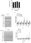
BDB induces autophagosome formation in DPCs: (a) Formation of autophagic vacuoles altered by BDB treatment for 24 h. The DPCs were stained with Cyto-ID fluorescence dye for 30 min at room temperature in the dark. Data were analyzed using a FACStar flow cytometer. (b) Changes in the expression levels of autophagy-related proteins in DPCs treated with BDB or minoxidil for 24 h. (c) Quantitative graph of changes in expression levels of autophagy-related proteins after treatment with BDB or minoxidil. (d) Changes in autophagy-related protein levels in DPCs treated with BDB for 0–24 h. (e) Quantitative graphs of changes in expression levels of autophagy-related proteins after BDB treatment for 0–24 h. Each dot indicates an independent intensity of the protein level. Data are presented as mean ± SD. * p < 0.05, ** p < 0.01, *** p < 0.001 versus the vehicle (DMSO)-treated control group.
Figure 5
Figure 5
BDB inhibits TGF-β1-induced activation of Smad2 in DPCs: (a) Changes in the nuclear/cytoplasmic translocation of Smad2/3 in DPCs treated with BDB or minoxidil for 24 h. (b) Quantitative graph of the changes in nuclear/cytoplasmic translocation of Smad2/3 after treatment with BDB or minoxidil. (c) Changes in the activation of Smad2 and Samd3 in DPCs treated with BDB and/or TGF-β1. (d) Quantitative graph of the changes in activation of Smad2 and Samd3 after treatment with BDB and/or TGF-β1. Each dot indicates an independent intensity of the protein levels. Data are presented as mean ± SD. ** p < 0.01, *** p < 0.001 compared with the control. ††† p < 0.001 compared with the TGF-β1-treated group.
Figure 5
Figure 5
BDB inhibits TGF-β1-induced activation of Smad2 in DPCs: (a) Changes in the nuclear/cytoplasmic translocation of Smad2/3 in DPCs treated with BDB or minoxidil for 24 h. (b) Quantitative graph of the changes in nuclear/cytoplasmic translocation of Smad2/3 after treatment with BDB or minoxidil. (c) Changes in the activation of Smad2 and Samd3 in DPCs treated with BDB and/or TGF-β1. (d) Quantitative graph of the changes in activation of Smad2 and Samd3 after treatment with BDB and/or TGF-β1. Each dot indicates an independent intensity of the protein levels. Data are presented as mean ± SD. ** p < 0.01, *** p < 0.001 compared with the control. ††† p < 0.001 compared with the TGF-β1-treated group.

Similar articles

Cited by

References

    1. Paus R., Haslam I.S., Sharov A.A., Botchkarev V.A. Pathobiology of chemotherapy-induced hair loss. Lancet Oncol. 2013;14:e50–e59. doi: 10.1016/S1470-2045(12)70553-3. - DOI - PubMed
    1. Alkhalifah A., Alsantali A., Wang E., McElwee K.J., Shapiro J. Alopecia areata update: Part I. Clinical picture, histopathology, and pathogenesis. J. Am. Acad. Dermatol. 2010;62:177–188. doi: 10.1016/j.jaad.2009.10.032. - DOI - PubMed
    1. Kaufman K.D. Androgens and alopecia. Mol. Cell. Endocrinol. 2002;198:89–95. doi: 10.1016/S0303-7207(02)00372-6. - DOI - PubMed
    1. Springer K., Brown M., Stulberg D.L. Common hair loss disorders. Am. Fam. Physician. 2003;68:93–102. - PubMed
    1. Jaller J.A., MacQuhae F., Nichols A.J. Chapter 26-Clinical Trials and Hair Loss. In: Miteva M., editor. Alopecia. Elsevier; Amsterdam, The Netherlands: 2019. pp. 267–284. - DOI

LinkOut - more resources