Review
. 2022 Mar 24;23(7):3537.
doi: 10.3390/ijms23073537.

An Updated Model of the Divisome: Regulation of the Septal Peptidoglycan Synthesis Machinery by the Divisome

Affiliations
Review

An Updated Model of the Divisome: Regulation of the Septal Peptidoglycan Synthesis Machinery by the Divisome

Mohamed Attaibi et al. Int J Mol Sci. .

Abstract

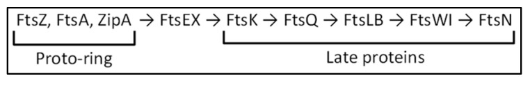
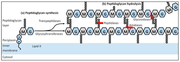
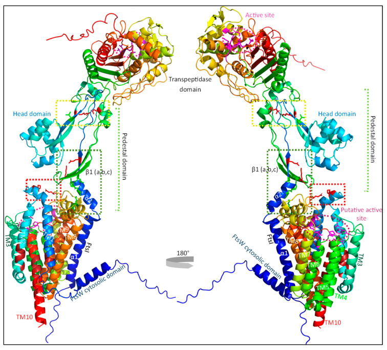
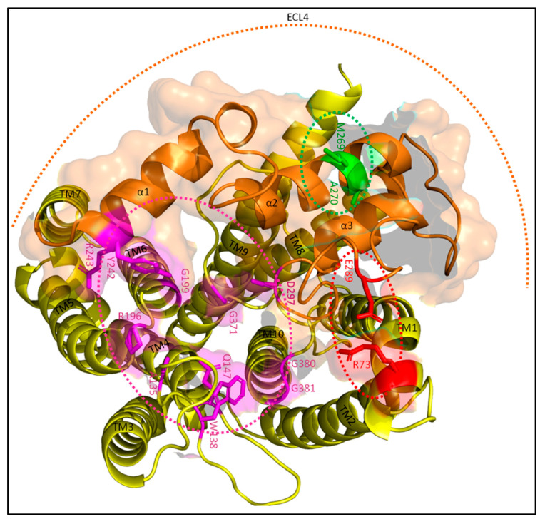
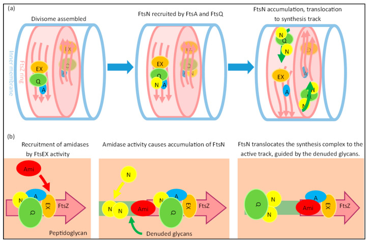
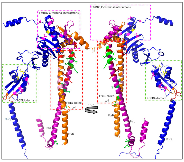
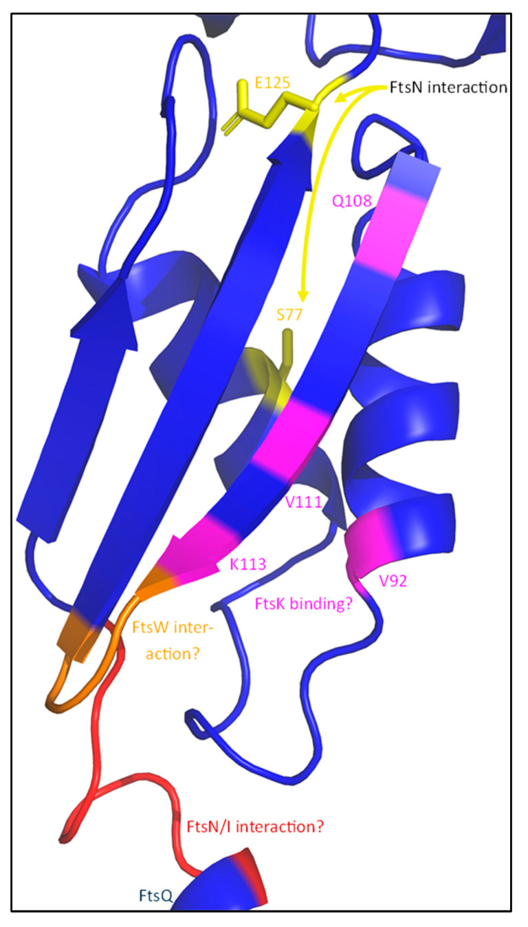
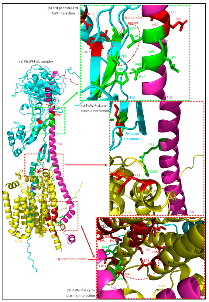
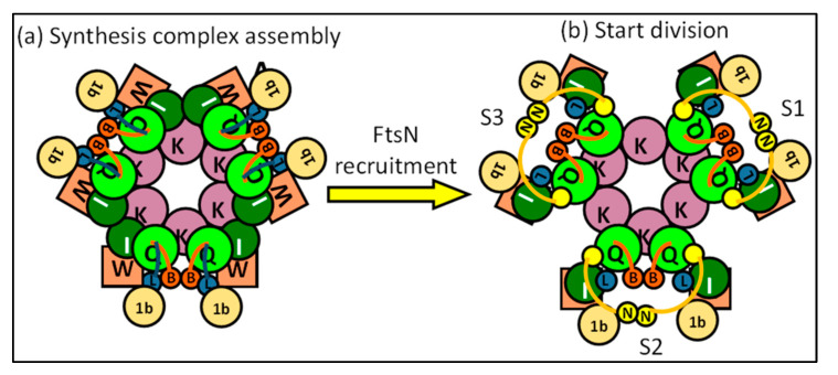
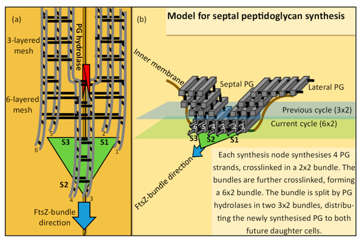
The synthesis of a peptidoglycan septum is a fundamental part of bacterial fission and is driven by a multiprotein dynamic complex called the divisome. FtsW and FtsI are essential proteins that synthesize the peptidoglycan septum and are controlled by the regulatory FtsBLQ subcomplex and the activator FtsN. However, their mode of regulation has not yet been uncovered in detail. Understanding this process in detail may enable the development of new compounds to combat the rise in antibiotic resistance. In this review, recent data on the regulation of septal peptidoglycan synthesis is summarized and discussed. Based on structural models and the collected data, multiple putative interactions within FtsWI and with regulators are uncovered. This elaborates on and supports an earlier proposed model that describes active and inactive conformations of the septal peptidoglycan synthesis complex that are stabilized by these interactions. Furthermore, a new model on the spatial organization of the newly synthesized peptidoglycan and the synthesis complex is presented. Overall, the updated model proposes a balance between several allosteric interactions that determine the state of septal peptidoglycan synthesis.

Keywords: FtsBLQ; FtsEX; FtsN; FtsWI; FtsZ; divisome; interactions; peptidoglycan hydrolases; regulation; septal peptidoglycan.


Conflict of interest statement

The authors declare no conflict of interest.

Figures

Figure 1
Figure 1
Hierarchical recruitment of divisome proteins.
Figure 2
Figure 2
Peptidoglycan biosynthesis and remodeling. This simplified scheme describes the steps needed to synthesize new peptidoglycan from precursor Lipid II that is synthesized in the cytoplasm. Lipid II is then flipped to the periplasmic side of the inner membrane by a flippase, where the synthesis of peptidoglycan occurs. (a) Lipid II is incorporated in an already growing peptidoglycan strand by glycosyltransferases. Adjacent Peptidoglycan strands are crosslinked by transpeptidases through the pentapeptides attached to the NAM residues, resulting in a peptidoglycan mesh. (b) Factors involved in the remodeling of the peptidoglycan mesh are PG hydrolases, which can be divided in multiple groups based on their targets. Peptidases cleave amide bonds in the cross-linked peptide chains, amidases cleave off the peptide chains attached to NAM residues and glycosidases that hydrolyze the glycosidic linkage between adjacent NAG and NAM residues.
Figure 3
Figure 3
The FtsWI peptidoglycan synthesizing holoenzyme. FtsI consists of a short cytoplasmic domain, a transmembrane domain (α1), a pedestal domain (including head domain) and a C-terminal transpeptidase domain. FtsW comprises a collection of 10 transmembrane domains (TM1-10) which are connected by periplasmic and cytoplasmic loops. Multiple regions that control the activity of FtsWI are indicated by the colored boxes and detailed further in Figure 3 and Figure 4. Although no crystal structure of the FtsWI is available, predictions of the structure have been produced with AlphaFold2 advanced and visualized with the PyMOL Molecular Graphics System, Version 2.5.2 Schrödinger, LLC. Sequences for the prediction were obtained from the UniProtKB database (P0ABG4-1 for FtsW and P0AD68-1 for FtsI).
Figure 4
Figure 4
FtsW active site. A single FtsW protein (yellow) is shown from above in the periplasm. The active site is indicated by the magenta–dotted ellipse with active site residues also highlighted in magenta. The ECL4 (orange) contains the putative catalytic residue D297, a region around M269 (green) that activates FtsW, and another region around E289 (red) that inhibits FtsW. E289 forms a putative interaction with R73 in this prediction. α1, α2 and α3 indicate the three α-helixes of ECL4. The FtsI interaction interface is highlighted in transparent orange. The active site residues are from the paper of Li et al. [67].
Figure 5
Figure 5
FtsI pedestal domain interaction with FtsW ECL4. FtsI (cyan) and FtsW (yellow) interact in the periplasm leading to the activation of FtsW. The membrane proximal part of the FtsI pedestal domain interacts with ECL4 of FtsW (orange). Activating (green) and suppressing (red) residues can be found in this region, and likely lead to stabilization of the active residue FtsWD297 in the active site. The proximal part of the pedestal domain contains multiple residues divided over an α-helix and β-sheet that activate FtsW. These include residues G57, S61 and L62 located on α2, and residue R210 on β1. Residue K211, located adjacent to R210, inhibits FtsW activation, likely through an interaction with Q65. The pedestal domain interacts mostly with the FtsW region around M269, which activates GTase activity. A cluster of residues at the end of the pedestal domain is important for the stability of this domain and appears to be involved in the regulation of the FtsI-mediated activation of FtsW. Residue R167, proposed to interact with E193, stabilizes the domain and thus inhibits FtsW activation, though it may also affect the activity of the FtsI TPase domain.
Figure 6
Figure 6
Intrinsic FtsWI regulation. FtsWI appear to have an intrinsic regulatory mechanism, which is expected to be affected by regulators. In a simplified scheme of the periplasmic side of FtsW (a), the catalytic residue D297 is located at the periphery of the active side (purple–dotted ring) in the loop between TM7 and TM8 that also contains the inhibiting E289 residue and the activating M269 and A270 residues. A balance between these two regions on the same loop appear to regulate FtsW GTase activity. A putative interaction between E289 and R73, located on TM1, likely destabilizes or sequesters D297 from the rest of the active site, leading to an inactive conformation. The hydrophobic region around M269 and A270 may antagonize this and lead to the stable positioning of D297 in the active site. (b) A speculative model describing the possible activation of FtsW by FtsI is shown. Regions of the pedestal domain of FtsI may interact with the α-helix containing M269 and A270, pushing the regulatory loop in an active conformation.
Figure 7
Figure 7
FtsA structure. FtsA in a cytoplasmic protein that binds to the inner membrane and acts as a membrane anchor for FtsZ. FtsA is divided into four domains which interact with different divisome components [81]. The 1A domain (green) is involved in membrane binding and has also been linked to FtsK binding (see text). The 2B domain (blue) contains a region that binds FtsZ and, together with the 1A and 2A domains, forms a nucleotide (ATP)-binding pocket at the FtsA core. The 1C domain (yellow) is involved in the recruitment of FtsN. FtsA oligomerizes through 1A–1C and 2A–2B interactions. Multiple SF mutants are shown in red, which are mostly located at the oligomerization interfaces.
Figure 8
Figure 8
FtsN structure. FtsN is a long bitopic protein, with a small cytoplasmic domain that interacts with FtsA (red), a transmembrane domain (orange), and a large periplasmic domain. The periplasmic domain is mostly unstructured, ending in a SPOR domain that binds to denuded glycans. The region in between the membrane and the SPOR domain contains three α-helixes, including an essential domain of 18 residues (green: EFtsN domain) partly located on the first helix.
Figure 9
Figure 9
FtsN active track formation. (a) A simplified model describing the possible formation of the FtsN-associated active track is shown. When the divisome is completely assembled, FtsN (N; yellow) can be recruited to the division site. FtsN is likely recruited by direct interactions with FtsA (A; blue) and FtsQ (Q; green). FtsEX (EX; orange) ATPase activity of FtsEX leads to the recruitment of amidases (Ami; red), which form denuded glycans. The SPOR domain of FtsN binds to the denuded glycans, leading to the accumulation of FtsN at the septum. FtsN, together with the synthesis complex represented by FtsQ, then translocates to the slower, active track away from the faster FtsZ-associated track. (b) The active track is proposed to be ‘carved’ out in the septal peptidoglycan by the amidases activated by FtsEX, which tightly interact with FtsZ from its recruitment. This mechanism describes how the presumed translocation of the synthesis complex may occur. This links the FtsN-track and FtsZ-track spatially, in line with experimental data. The pink arrows indicate the dynamics of FtsZ filament bundles, and the green arrows at the top indicate the dynamics of the active FtsN-associated track.
Figure 10
Figure 10
The FtsBLQ regulatory subcomplex. Shown is the FtsBLQ subcomplex responsible for the regulation of the septal peptidoglycan synthesis machinery. The largest subunit, FtsQ, acts as a scaffold and comprises a small cytosolic domain, a transmembrane domain, and a large periplasmic domain. FtsB and FtsL have a similar structure, mostly consisting of a long helical structure, and form a coiled coil together. FtsB has a short cytoplasmic tail, while FtsL carries a large cytosolic double helical structure involved in the recruitment of FtsW. Regions or interactions that are important for sPG synthesis regulation are highlighted by the colored boxes and are further detailed in Figure 11 (POTRA domain), Figure 13 (FtsBL coiled coil) and Figure 15 (FtsBLQ C-terminal interactions). The structures shown here are predictions produced with AlphaFold2, advanced and visualized with the PyMOL Molecular Graphics System, Version 2.5.2 Schrödinger, LLC. Sequences for the prediction were obtained from the UniProtKB database (P06136-1 for FtsQ, Q9HXZ6-1 for FtsB and P0AEN4- for FtsL).
Figure 11
Figure 11
The FtsQ POTRA domain. FtsQ interacts with multiple divisome proteins through its POTRA domain, which is part of the membrane-proximal periplasmic domain. A loop between the TM domain and the first strand of the β-sheet (50–57 in red) is necessary for the interaction with FtsI (and FtsN). Additional FtsN interacting residues are shown in yellow (S77, E125). The POTRA domain also contains a FtsK-binding site (purple), which includes a hydrophobic pocket (V92, V111, L57). Furthermore, a FtsW interaction interface is expected somewhere in the region between Q108 and E125. This is likely the loop between the two strands of the β-sheet, as this is the region predicted to be closest to the membrane-embedded FtsW (orange).
Figure 12
Figure 12
Interactions between FtsL and FtsWI. FtsL interacts with multiple regions of FtsWI, which likely leads to a stable active conformation. (a) The complex formed between FtsW (yellow), FtsI (cyan) and FtsL (purple) is shown. (b) A periplasmic interaction between the FtsL AWI domain (green) and the pedestal domain of FtsI is predicted to be important for FtsWI activation. A hydrophobic pocket around FtsI residues V84 and V86 (red dotted ellipse) appears to interact with FtsL residue L86. (c) FtsL also appears to interact with FtsW close to the IM on the periplasmic side. A hydrogen bond between FtsL residue R61 and FtsW residue G282 is formed in the predicted structure. The R61–G282 interaction (green) may stabilize the periplasmic loop in a more active conformation. (d) FtsL recruits FtsW through its cytoplasmic domain and this interaction also plays a minor role in FtsW activation. A hydrophobic pocket formed by residues om TM2 and TM10 of FtsW (brick-red) interacts with the FtsL cytoplasmic domain (L24, I28; green), possibly stabilizing the TM domains that carry active site-residues at the periplasmic side. The structures shown here are predictions produced with AlphaFold2 advanced, and visualized with PyMOL Molecular Graphics System, Version 2.5.2 Schrödinger, LLC. Sequences for the prediction were obtained from the UniProtKB database (P0AEN4-1 for FtsL, P0ABG4-1 for FtsW, P0AD68-1 for FtsI).
Figure 13
Figure 13
FtsBL coiled coil stability. Modulating the stability of the FtsBL coiled coil structure is an integral part of sPG synthesis regulation. The stability is dynamically affected by polar (Q39, R67, R74; N50) and hydrophobic (L46, L77, W81) clusters located at the inner part of the coiled coil between the transmembrane and the CCD/AWI domains. FtsB contains an alanine-rich patch at the other face of the helix, forming a dimerization/intercoil domain. The CCD (red) and AWI (green) domains just upstream are involved in the suppression and activation of sPG synthesis, respectively. Just above the FtsB CCD domain, a region stably interacting with FtsQ can be found.
Figure 14
Figure 14
Potential FtsQ–FtsI interactions. The potential FtsQ–FtsI interaction interface described in Table 2 is shown here in the predicted structures of FtsI and FtsQ. The FtsI region forms a helix, while the FtsQ region is an unstructured loop. Possible regulating interactions have been indicated with the red lines and the FtsW-activating residues of FtsI in green.
Figure 15
Figure 15
FtsBLQ C-terminal interaction. The C-terminal domain of FtsQ interacts with FtsB, through the formation of a multi-stranded β-sheet (βL, βB and βQ). Stability in this domain is important for the interactions, as mutating the residues marked in purple has a negative effect on FtsQ–FtsB interaction. Furthermore, a C-terminal interaction between FtsB and FtsL is predicted, as FtsL forms a β-strand interacting with the FtsB–FtsQ β-sheet (βL). This may be stabilized further by FtsQ. An additional FtsN-binding residue can be found in this region of FtsQ (S242; yellow) which is vital for FtsN–FtsQ interaction. This FtsN interaction may induce instability in this region, thus changing the conformation of FtsBL to an activation one.
Figure 16
Figure 16
Regulation of FtsWI–PBP1b by FtsBLQ and FtsN. A model is proposed where FtsBLQ and FtsN stabilize the FtsWI–PBP1b septal peptidoglycan synthesis machinery in active or inactive conformations based on certain allosteric interactions. These interactions are numbered and either suppress (red), increase (green), or are neutral (black) regarding sPG synthesis. (a) FtsW/L cytoplasmic interaction. This interaction between FtsL and FtsW in the cytoplasm recruits the synthesis machinery to the division site. While primarily involved in recruitment, the interaction may also be involved in the stabilization of FtsW in a more active conformation. (b) FtsQ–FtsWI. The membrane proximal part of the FtsQ POTRA domain seems to interact with a periplasmic loop of FtsW (between TM1 and TM2) and the pedestal domain of FtsI. (c) C-terminal FtsBLQ interaction. A strong periplasmic interaction between FtsB and FtsQ is important for the stability of the FtsBL coiled coil structure, as well as an additional C-terminal FtsB–FtsL interaction that occurs in the same region. This increased stability is thought to stabilize the whole complex in an inactive conformation, thus negatively affecting sPG synthesis. (d) PBP1b–FtsL. A direct interaction between PBP1b and the CCD domain of FtsL (red) suppresses PBP1b GTase activity. (e) PBP1b–FtsN. FtsN (yellow) also directly interacts with the same PBP1b region, outcompetes FtsL–CCD, and leads to the activation of PBP1b GTase activity. (f) FtsN–FtsQ. FtsN is proposed to interact with the same FtsQ region as FtsI and leads to dissociation of the inhibitory FtsQ–FtsWI interaction. (g) FtsL–FtsI. An interaction between the now available FtsL–AWI domain (green) and the pedestal domain of FtsI stabilizes FtsWI in a highly active conformation, starting septal peptidoglycan synthesis. (h) C-terminal FtsBLQ (in)stability. An interaction between FtsN and the C-terminal domain of FtsQ (around residue S242) is proposed to lead to instability in the C-terminal FtsBLQ interaction through a conformational change in FtsQ. Together with interactions (e,f), the instability in the FtsBL coiled coil domain is increased, which leads to dissociation of the upper part of the coiled coil and subsequently to the activating FtsL–FtsI interaction (see interaction (g)). (i) FtsN SPOR interacting with denuded glycans. The SPOR domain of FtsN binds to denuded glycan as shown, and most likely directs the synthesis complex to ‘open’ PG, where synthesis can occur. New PG strands are formed when the whole synthesis complex is in its active state, induced by the sum of activating interactions (green). This model is not on scale and does not represent the possible stoichiometry of a complete synthesis node. M and G represent NAM and NAG residues, respectively.
Figure 17
Figure 17
Stoichiometry of the synthesis complex. (a) A synthesis complex, shown from above in the periplasm, is assembled around FtsK hexamers, consisting of three synthesis nodes. Each synthesis node is comprised of 2 FtsBLQ–FtsWI–PBP1b subcomplexes, which assemble around FtsK through FtsQ. (b) FtsN recruitment activates the synthesis complex, leading to sPG synthesis by the three synthesis nodes (S1, S2 and S3).
Figure 18
Figure 18
A speculative model describing peptidoglycan synthesis based on the synthesis complex. (a) At top-down scheme from the periplasm is shown. Here, the synthesis complex is in green and travels around the division site guided by FtsZ filament bundles (blue arrow). The three synthesis nodes (S1, S2, and S3) each produce a PG bundle consisting of 4 PG strands in a 2 × 2 conformation (grey lines = NAM-NAG polymers; black lines = crosslinked peptides). The numbers (1, 2, 3, 4, 5, and 6) indicate the two PG strands, vertically crosslinked in the model, which are further crosslinked with two PG strands synthesized by the other FtsWI–PBP1b subcomplex localized in the synthesis node. Further crosslinking of the newly synthesized PG bundles leads to a PG mesh of 6 strands wide and 2 strands high (6-layered mesh). The 6-strand-wide PG mesh is speculated to be divided by lagging PG hydrolases in two separate meshes, each 3 strands wide and 2 strands high (3-layered mesh). The two 3 × 2 PG meshes would be divided over two future daughter cells by the outer membrane, which would be in line with the current view concerning the structural conformation of PG at the cell poles. (b) We see the same speculative model describing septal peptidoglycan synthesis from a frontal view. Here, sPG synthesis is shown from a frontal perspective. The inner membrane (brown line) and the PG layer (grey blocks = NAM-NAG polymers; black lines = crosslinked peptides) are shown, while the outer membrane is excluded for simplicity. A distinction is made between lateral PG, which is mostly single-layered, while septal PG is three-layered in the model. The synthesis complex (green) produces the 6 × 2 PG mesh, which is not yet separated by PG hydrolases (not shown here) in the current cycle of remodeling. The PG mesh produced in the previous cycle has already been separated in two 3 × 2 meshes, where each mesh is distributed over a future daughter cell. This model, based on the supposed conformation of the synthesis complex, provides a cycle-based mechanism, where during each cycle a new PG mesh is produced inward from the PG produced during the previous cycle (or from a lateral, single-layered PG). The separation by PG hydrolases during the previous cycle may lead to denuded glycans, which can be bound by FtsN, providing a platform for the synthesis complex to produce new PG in an inward manner.

References

    1. Angert E.R. Alternatives to binary fission in bacteria. Nat. Rev. Microbiol. 2005;3:214–224. doi: 10.1038/nrmicro1096. - DOI - PubMed
    1. den Blaauwen T., Hamoen L.W., Levin P.A. The divisome at 25: The road ahead. Curr. Opin. Microbiol. 2017;36:85–94. doi: 10.1016/j.mib.2017.01.007. - DOI - PMC - PubMed
    1. Aarsman M.E.G., Piette A., Fraipont C., Vinkenvleugel T.M.F., Nguyen-Distèche M., Den Blaauwen T. Maturation of the Escherichia coli divisome occurs in two steps. Mol. Microbiol. 2005;55:1631–1645. doi: 10.1111/J.1365-2958.2005.04502.X. - DOI - PubMed
    1. Gamba P., Veening J.W., Saunders N.J., Hamoen L.W., Daniel R.A. Two-step assembly dynamics of the Bacillus subtilis divisome. J. Bacteriol. 2009;191:4186–4194. doi: 10.1128/JB.01758-08. - DOI - PMC - PubMed
    1. Du S., Henke W., Pichoff S., Lutkenhaus J. How FtsEX localizes to the Z ring and interacts with FtsA to regulate cell division. Mol. Microbiol. 2019;112:881–895. doi: 10.1111/MMI.14324. - DOI - PMC - PubMed

MeSH terms

LinkOut - more resources