. 2022 Mar 25;23(7):3617.
doi: 10.3390/ijms23073617.

TRPV2: A Key Player in Myelination Disorders of the Central Nervous System

Affiliations

TRPV2: A Key Player in Myelination Disorders of the Central Nervous System

Jennifer Enrich-Bengoa et al. Int J Mol Sci. .

Abstract

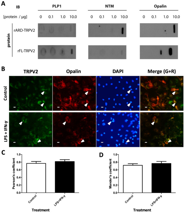
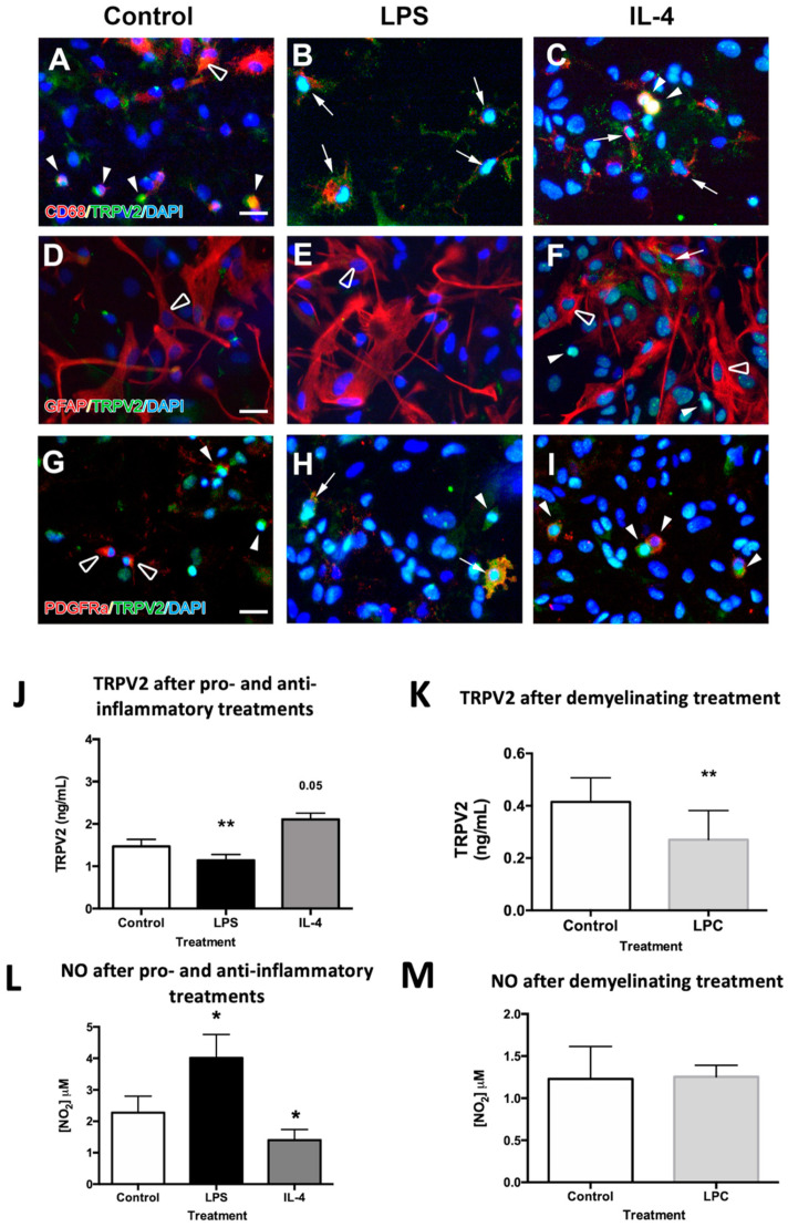
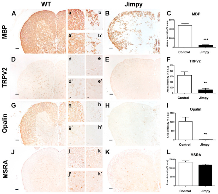
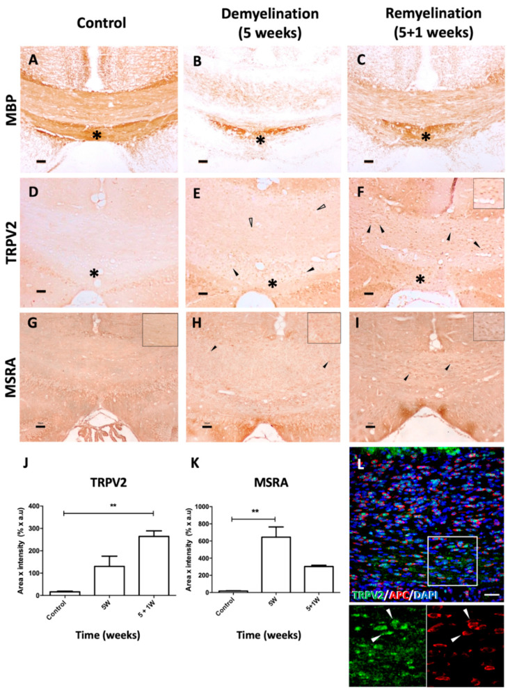
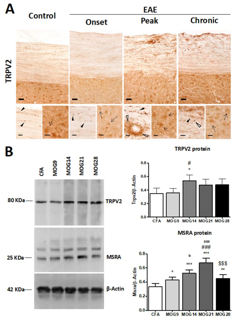
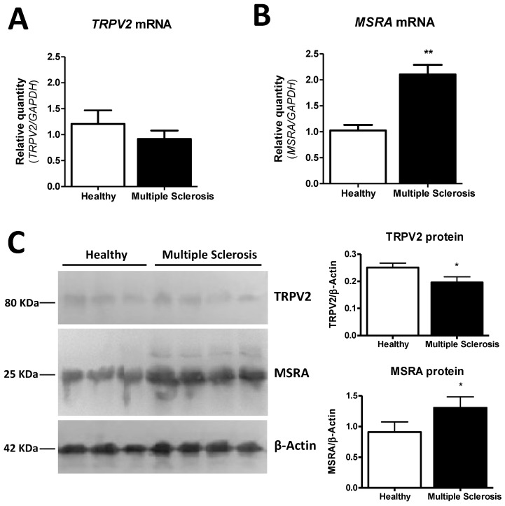
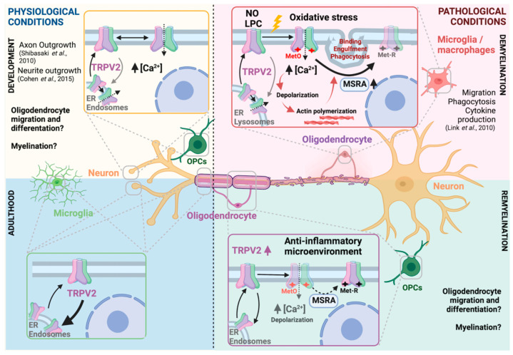
Transient potential receptor vanilloid 2 (TRPV2) is widely expressed through the nervous system and specifically found in neuronal subpopulations and some glial cells. TRPV2 is known to be sensitized by methionine oxidation, which results from inflammation. Here we aim to characterize the expression and regulation of TRPV2 in myelination pathologies, such as hypomyelination and demyelination. We validated the interaction between TRPV2 and its putative interactor Opalin, an oligodendrocyte marker, in mixed glial cultures under pro- and anti-inflammatory conditions. Then, we characterized TRPV2 time-course expression in experimental animal models of hypomyelination (jimpy mice) and de-/remyelination (cuprizone intoxication and experimental autoimmune encephalomyelitis (EAE)). TRPV2 showed upregulation associated with remyelination, inflammation in cuprizone and EAE models, and downregulation in hypomyelinated jimpy mice. TRPV2 expression was altered in human samples of multiple sclerosis (MS) patients. Additionally, we analyzed the expression of methionine sulfoxide reductase A (MSRA), an enzyme that reduces oxidated methionines in TRPV2, which we found increased in inflammatory conditions. These results suggest that TRPV2 may be a key player in myelination in accordance with the recapitulation hypothesis, and that it may become an interesting clinical target in the treatment of demyelination disorders.

Keywords: Opalin; multiple sclerosis; myelination; oxidative stress; recapitulation theory; transient potential receptor vanilloid 2.


Conflict of interest statement

The authors declare no conflict of interest.

Figures

Figure 1
Figure 1
TRPV2 interaction with Opalin, NTM and PLP1 in vitro during basal and inflammatory conditions. (A) Rat TRPV2 FL protein and rat ARD of TRPV2 were immobilized on membranes and incubated with increasing concentrations of rat protein brain lysates (0–10 µg). Opalin, NTM and PLP1 were detected by immunoblot. (B) Immunocytochemistry in mouse mixed glial primary cultures shows TRPV2 (green), Opalin (red) and DAPI (blue). TRPV2-Opalin colocalization is shown as yellow signal (merge) and both proteins are co-expressed in some cells (arrowheads). Expression of these proteins is mainly found in the membrane. (C) TRPV2–Opalin colocalization expressed as Pearson’s coefficient in basal and inflammatory conditions. Both proteins show a high degree of colocalization. (D) TRPV2 colocalization with Opalin, expressed as Mander’s coefficient M2 (fraction of TRPV2 signal overlapping Opalin) in basal and inflammatory conditions. Both proteins show a high degree of colocalization. Results were expressed as mean ± SEM. Statistical analysis of the results were performed with an unpaired Student’s t-test (p < 0.05 is statistically significant). Scale bar = 20 µm.
Figure 2
Figure 2
TRPV2 expression in mixed glia cultures of mice after pro-inflammatory (LPS), anti-inflammatory (IL-4) and demyelinating (LPC) treatments. (AI) Double immunohistochemical staining allowed for the determination of whether TRPV2 was expressed in microglia (AC), astrocytes (DF) or oligodendrocyte cells (GI). TRPV2 was highly expressed in the cell body of some microglia (full arrowheads) but not all of them (empty arrowheads) in control conditions, and TRPV2 was spread afterwards through the cell body of activated microglia cells, with an expression pattern highly coincident with CD68+ lysosomes (arrows). While no expression of TRPV2 was found in astrocytes (empty arrowheads), OPCs showed either low or absent expression in control conditions (full and empty arrowheads, respectively) and increased expression throughout the cell body (arrows) and the principal soma (arrowheads) after both pro-inflammatory and anti-inflammatory treatments. (J,K) Quantification of TRPV2 protein (ng/mL) by ELISA in secondary mixed glial cultures in control conditions, and after LPS and IL-4 treatments (J) and in quaternary mixed glial cultures, containing myelin-binding protein (MBP)+ oligodendrocytes, in demyelinating conditions (K). Determination of TRPV2 showed a significant decrease in TRPV2 after LPS and LPC treatments (J,K), and an increase after IL-4 treatment (p = 0.05) compared to control conditions (J). (L,M) Quantification of NO secondary mixed glial cultures in control conditions, and after LPS and IL-4 treatments (L) and in quaternary mixed glial cultures in demyelinating conditions (M). An increase in NO concentration was observed in cell cultures after LPS treatment compared to control conditions, while a significant decrease was observed after IL-4 treatment. After LPC treatment, NO remained stable compared to the control. Statistical analysis was performed by one-way ANOVA followed by Dunnett’s multiple comparison compared to the control in the LPS and IL-4 treated cultures (* p < 0.05, ** p < 0.01), and a paired Student’s t-test for LPC treatment (** p < 0.01). Scale bar (AI) = 25 µm.
Figure 3
Figure 3
TRPV2, Opalin and MSRA expression in spinal cord of wild-type (WT) and hypomyelination jimpy mice. Immunohistochemical staining against MBP (A,B), TRPV2 (D,E), Opalin (G,H) and MSRA (J,K) were performed in spinal cord sections of 21-day-old WT and jimpy mice, and immunoreactivity was quantified and analyzed as Area x Intensity. Results show that MBP was importantly reduced in both GM (a,b) and WM (a’,b’) of jimpy mice compared to WT (AC). TRPV2 expression was mainly located in neurons in GM (d’) and glial cells, putatively oligodendrocytes, and in WM (d) of WT. A significant decrease in TRPV2 expression in the spinal cord of jimpy mice was observed compared to WT (E,F,e,e’). Opalin expression was importantly found in WM of WT mice (G,g), and to a lesser extent also in GM (g’), but showed a marked decrease in WM and GM of jimpy mice (HI,h,h’). MSRA was the only molecule that did not suffer an important reduction of its expression in both WM (j,k) and GM (j’,k’) in jimpy mice (L). Results were expressed as mean ± SEM. Statistical analysis of the results were performed with an unpaired Student’s t-test (** p < 0.01, *** p < 0.001). Scale bar (AK) = 50 µm; (ak,a’k’) = 20 µm.
Figure 4
Figure 4
TRPV2 and MSRA expression in WM of cuprizone-induced demyelination and remyelination in mice. (AI) Representative images of immunohistochemical staining directed against MBP, TRPV2 and MSRA in the CC of control mice (A,D,G) and cuprizone-intoxicated mice after demyelination (B,E,H, 5 weeks of treatment) and during remyelination (C,F,I, 5 weeks of treatment + 1 week of normal diet). (AC) MBP immunostaining shows the myelinated CC of WT mice in basal conditions (A). At 5 weeks of cuprizone treatment, the CC is largely demyelinated (B) with a myelination-resistant area (*), and after one week of normal diet, the CC is mostly remyelinated (C). Control mice show undetectable levels of TRPV2 and MSRA in the CC (D and G). After demyelination, variably low and high levels of TRPV2 were observed in cell bodies (empty arrowheads and arrowheads, respectively in E,F). High levels of TRPV2 were clearly observed at remyelination (inset in F). During remyelination, most TRPV2+ cells co-expressed APC (full arrowheads in L), a marker for mature oligodendrocytes. In the CC area resistant to demyelination (* in E,F), TRPV2+ cells showed high levels of expression in both conditions. An increase in MSRA levels of expression were also observed in cell bodies after demyelination and remyelination (arrowheads in H and I). In demyelination, most cells expressed low levels of MSRA in the CC (inset in H), while during remyelination, a lower number of MSRA+ cells expressing higher levels of MSRA were observed (inset in I). (JK) Quantification of TRPV2 and MSRA expression after cuprizone treatment. (J) Quantitative analysis of TRPV2 immunoreactivity in the CC confirmed the progressive increase of TRPV2 expression in demyelinating and remyelinating conditions compared to control mice, which was statistically significant in the latter. (K) Quantitative analysis of MSRA immunoreactivity in the CC showed that the increase of MSRA expression during demyelination was statistically significant when compared to control mice. Data are shown as ± SEM. Statistical analysis was performed by one-way ANOVA followed by Tukey’s multiple comparison test (** p < 0.01). Scale bar (AI) = 50 μm; (L) = 100 μm.
Figure 5
Figure 5
TRPV2 expression in spinal cord samples from WT and EAE mice. (A) Immunohistochemical staining of TRPV2 in spinal cord of control and EAE-induced mice in the onset, peak and in the chronic phase, during remission. In control conditions, TRPV2 is expressed in neurons in GM (inset, arrows), and in oligodendrocytes in WM (inset, arrowheads). After EAE induction, an increase in TRPV2 expression is mainly located in inflammatory focuses at the peak clinical symptomatology in WM (empty arrowheads). (B) MSRA and TRPV2 protein expression in thoracic spinal cord samples from CFA and MOG mice (9, 14, 21 and 28 days post-immunization). (B) A significant increase in TRPV2 protein levels (#, p = 0.0014) is determined in MOG14 mice when compared with CFA (p < 0.05) and when compared with MOG9 (p < 0.05). A significant increase (p < 0.05) in MSRA protein levels is observed in all groups of mice treated with MOG when compared with CFA mice, as well as between different groups treated with MOG: MOG9 vs. MOG21 (###, p < 0.001), MOG9 vs. MOG14 (b, p < 0.05), MOG21 vs. MOG28 ($$$, p < 0.001), MOG14 vs. MOG21 (aaa, p < 0.001). Bars show mean ± SEM; * p < 0.05, ** p < 0.01 and *** p < 0.001 vs. CFA using one-way ANOVA and Newman–Keuls post-test. Scale bars = 50 μm; (insets) = 20 μm.
Figure 6
Figure 6
MSRA and TRPV2 expression in frontal cortex samples from healthy subjects (n = 4) and MS (n = 6) patients. (A) TRPV2 mRNA (p = 0.3314) is not changed between MS and healthy samples. (B) A significant upregulation in MSRA mRNA (p = 0.0014) is found in MS; (C) A significant decrease in TRPV2 protein levels (p = 0.0232) and a significant increase in MSRA protein levels (p = 0.0124) were determined in MS samples by western blot. Bars show mean ± SEM; * p < 0.05, ** p < 0.01 using unpaired Student’s t-test.
Figure 7
Figure 7
TRPV2-centered cellular cross-talk model in physiological and pathophysiological myelination. During embryonic development, TRPV2 develops an essential role in neurite and axon outgrowth. In that period, TRPV2 is known to be expressed and activated in neurons, and putatively in OPCs, where possible cross-talk between both cells might be triggered through TRPV2 activation. In adulthood, TRPV2 expression is found in microglia, neurons and oligodendrocytes. Upon an insult such as demyelination, TRPV2, as a non-selective ion channel, is activated. Some known triggering stimuli are NO, LPC or oxidative stress, the latter causing methionine oxidation which facilitates TRPV2 sensitization. TRPV2 activation promotes a sodium/calcium inward influx from extracellular media and/or ER or endosomes. Consequently, cells are depolarized, which initiate processes such as actin polymerization or phagocytosis in microglia or innate immune cells. In those cells, TRPV2 has been involved in migration, phagocytosis or cytokine production. TRPV2 inactivation is promoted by MSRA enzyme, by reducing oxidized methionine. In our results, we observed both an increase of TRPV2 and MSRA in demyelinating scenarios. During remyelination, OPCs proliferate and migrate to the demyelinated area, where they differentiate through several stages to mature oligodendrocytes, and start myelination. Our results showed increased TRPV2 expression during this period. Overall, ubiquitous TRPV2 expression and activation in de- and remyelinating processes suggest a multicellular cross-talk that promotes myelin repair at all levels. TRPV2 is relevant in development and demyelination, making this model suitable for the recapitulation hypotheses. Created with BioRender.com (accessed on 10 January 2022) [8,9,10].

Similar articles

Cited by

References

    1. Perálvarez-Marín A., Doñate-Macian P., Gaudet R. What do we know about the transient receptor potential vanilloid 2 (TRPV2) ion channel? FEBS J. 2013;280:5471–5487. doi: 10.1111/febs.12302. - DOI - PMC - PubMed
    1. Fricke T.C., Echtermeyer F., Zielke J., de la Roche J., Filipovic M.R., Claverol S., Herzog C., Tominaga M., Pumroy R.A., Moiseenkova-Bell V.Y., et al. Oxidation of methionine residues activates the high-threshold heat-sensitive ion channel TRPV2. Proc. Natl. Acad. Sci. USA. 2019;116:24359–24365. doi: 10.1073/pnas.1904332116. - DOI - PMC - PubMed
    1. Kojima I., Nagasawa M. TRPV2. Handb. Exp. Pharmacol. 2014;222:247–272. doi: 10.1007/978-3-642-54215-2_10. - DOI - PubMed
    1. Caterina M.J., Rosen T.A., Tominaga M., Brake A.J., Julius D. A capsaicin-receptor homologue with a high threshold for noxious heat. Nature. 1999;398:436–441. doi: 10.1038/18906. - DOI - PubMed
    1. Maksoud M.J.E., Tellios V., An D., Xiang Y., Lu W. Nitric oxide upregulates microglia phagocytosis and increases transient receptor potential vanilloid type 2 channel expression on the plasma membrane. Glia. 2019;67:2294–2311. doi: 10.1002/glia.23685. - DOI - PubMed