Review
. 2022 Mar 26;23(7):3627.
doi: 10.3390/ijms23073627.

Synapses: The Brain's Energy-Demanding Sites

Affiliations
Review

Synapses: The Brain's Energy-Demanding Sites

Andreia Faria-Pereira et al. Int J Mol Sci. .

Abstract

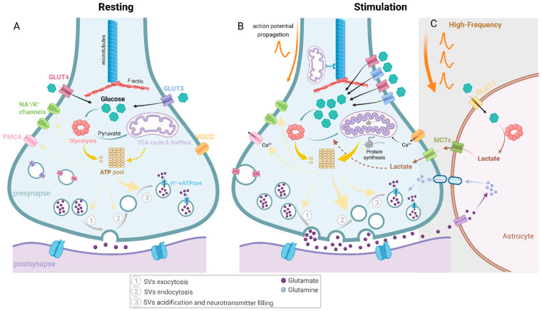
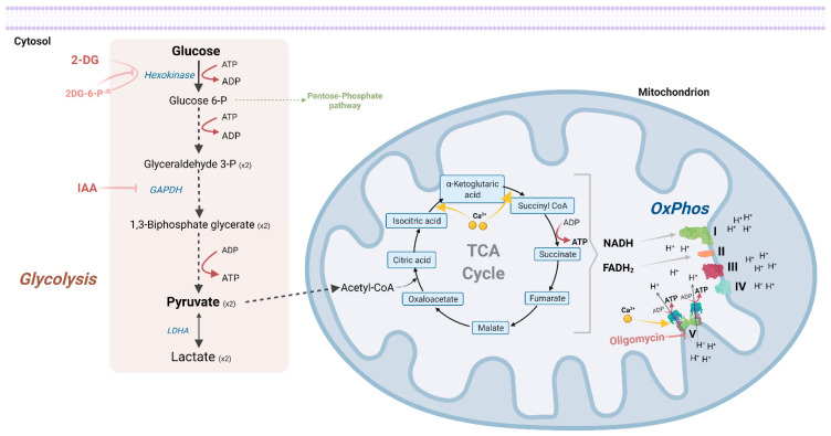
The brain is one of the most energy-consuming organs in the mammalian body, and synaptic transmission is one of the major contributors. To meet these energetic requirements, the brain primarily uses glucose, which can be metabolized through glycolysis and/or mitochondrial oxidative phosphorylation. The relevance of these two energy production pathways in fulfilling energy at presynaptic terminals has been the subject of recent studies. In this review, we dissect the balance of glycolysis and oxidative phosphorylation to meet synaptic energy demands in both resting and stimulation conditions. Besides ATP output needs, mitochondria at synapse are also important for calcium buffering and regulation of reactive oxygen species. These two mitochondrial-associated pathways, once hampered, impact negatively on neuronal homeostasis and synaptic activity. Therefore, as mitochondria assume a critical role in synaptic homeostasis, it is becoming evident that the synaptic mitochondria population possesses a distinct functional fingerprint compared to other brain mitochondria. Ultimately, dysregulation of synaptic bioenergetics through glycolytic and mitochondrial dysfunctions is increasingly implicated in neurodegenerative disorders, as one of the first hallmarks in several of these diseases are synaptic energy deficits, followed by synapse degeneration.

Keywords: brain energy metabolism; glycolysis; mitochondria; synapses.


Conflict of interest statement

The authors declare no conflict of interest.

Figures

Figure 2
Figure 2
The dynamic energy requirements and fulfilments of presynapses upon distinct activation states. Glucose is the main energy substrate in the brain and given that synapses are the most energy-demanding sites in the brain, glucose degradation is important to provide ATP for synaptic pathways. Glucose is imported through glucose transporters (GLUT3 and GLUT4) and can be degraded through glycolysis in the cytosol and generate pyruvate and, ultimately, lactate. Pyruvate can be imported to mitochondria where, through multiple reactions shown in Figure 1, it will generate higher yields of ATP but at a lower rate than glycolysis [6]. Multiple reactions at presynapses require ATP, namely, synaptic vesicle recycling, endocytosis, acidification and neurotransmitter filling, priming (not represented in this figure) and exocytosis. Additionally, several ion channels in the presynaptic membrane require ATP, including sodium-potassium voltage-gated channels (Na+/K+ channels) and plasma membrane Ca2+-ATPase (PMCA), necessary to extrude Ca2+ that accumulates inside presynapses. (A) In a resting state, minimal energy is necessary to fuel these membrane ion channels [39], and it is speculated that due to the lack of stimuli, the energetic burden of synaptic vesicles’ (SVs) endo- and exocytosis is also reduced, despite some spontaneous release. Recently, in rat hippocampal neurons, it was suggested that SV re-acidification (through H+-vATPases) was the major energy consumer in presynapses (~44%) [39]. (B) Upon synaptic stimulation, the demand for energy is increased. Although the energy proportion required for each synaptic ATP-dependent process is not yet defined, SV endocytosis is assumed to be one of the most energy-demanding processes. Both glycolysis and OxPhos have been shown to be important to meet the activity-driven energy requirements. In active synapses, mitochondria present a higher volume and an increased cristae density [44]. Additionally, translation of both mitochondrial [45]- and nuclear-encoded mitochondrial [46] proteins have been shown to be increased upon synaptic activity. Additionally, recruitment of axonal mitochondria has also been shown to be important upon synaptic energy stress [47]. (C) Despite the controversy of the astrocyte-neuron lactate shuttle (ANLS) hypothesis, astrocytic lactate has been important to maintain synaptic transmission under high-frequency stimuli (HFS) [23,48,49]. Under these conditions, augmented glutamate levels in the glutamatergic synaptic clefts might boost astrocytic glutamate uptake to reconvert it back to glutamine. Concomitantly, astrocytic glycolysis is increased, generating lactate, which will be delivered to neurons through monocarboxylate transporters (MCTs) present in both neuronal and astrocytic membranes. This lactate will complement neuronal glycolysis and OxPhos in fulfilling the high SV endocytosis resultant from HFS.
Figure 1
Figure 1
Glycolysis and oxidative phosphorylation pathways are the major ATP providers.

References

    1. Attwell D., Laughlin S.B. An Energy Budget for Signaling in the Grey Matter of the Brain. J. Cereb. Blood Flow Metab. 2001;21:1133–1145. doi: 10.1097/00004647-200110000-00001. - DOI - PubMed
    1. Harris J.J., Attwell D. The Energetics of CNS White Matter. J. Neurosci. 2012;32:356–371. doi: 10.1523/JNEUROSCI.3430-11.2012. - DOI - PMC - PubMed
    1. Howarth C., Peppiatt-Wildman C., Attwell D. The Energy Use Associated with Neural Computation in the Cerebellum. J. Cereb. Blood Flow Metab. 2009;30:403–414. doi: 10.1038/jcbfm.2009.231. - DOI - PMC - PubMed
    1. Harris J.J., Jolivet R., Attwell D. Synaptic Energy Use and Supply. Neuron. 2012;75:762–777. doi: 10.1016/j.neuron.2012.08.019. - DOI - PubMed
    1. Howarth C., Gleeson P., Attwell D. Updated Energy Budgets for Neural Computation in the Neocortex and Cerebellum. J. Cereb. Blood Flow Metab. 2012;32:1222–1232. doi: 10.1038/jcbfm.2012.35. - DOI - PMC - PubMed

LinkOut - more resources