. 2022 Mar 27;23(7):3674.
doi: 10.3390/ijms23073674.

Protective Effects of Mitochondrial Uncoupling Protein 2 against Aristolochic Acid I-Induced Toxicity in HK-2 Cells

Affiliations

Protective Effects of Mitochondrial Uncoupling Protein 2 against Aristolochic Acid I-Induced Toxicity in HK-2 Cells

Chen Feng et al. Int J Mol Sci. .

Abstract

Aristolochic acid I (AA I) is one of the most abundant and toxic aristolochic acids that is reported to cause Aristolochic acid nephropathy (AAN). This paper was designed to assess whether mitochondrial Uncoupling Protein 2 (UCP2), which plays an antioxidative and antiapoptotic role, could protect human renal proximal tubular epithelial (HK-2) cells from toxicity induced by AA I. In this study, HK-2 cells were treated with different concentrations of AA I with or without UCP2 inhibitor (genipin). To upregulate the expression of UCP2 in HK-2 cells, UCP2-DNA transfection was performed. The cell viability was evaluated by colorimetric method using MTT. A series of related biological events such as Reactive Oxygen Species (ROS), Glutathione peroxidase (GSH-Px), and Malondialdehyde (MDA) were evaluated. The results showed that the cytotoxicity of AA I with genipin group was much higher than that of AA I alone. Genipin dramatically boosted oxidative stress and exacerbated AA I-induced apoptosis. Furthermore, the increased expression of UCP2 can reduce the toxicity of AA I on HK-2 cells and upregulation of UCP2 expression can reduce AA I-induced oxidative stress and apoptosis. In conclusion, UCP2 might be a potential target for alleviating AA I-induced nephrotoxicity.

Keywords: apoptosis; aristolochic acid I (AA I); nephrotoxicity; oxidative stress; uncoupling protein 2 (UCP2).


Conflict of interest statement

The authors declare no conflict of interest.

Figures

Figure 1
Figure 1
(A) Aristolochiaceae family and the chemical structure of main Aristolochic acids components AA I. (B) Toxicity of AA I to HK-2 cells. MTT was used for cell viability and cytotoxicity. HK-2 cells were treated with different concentrations of AA I with or without genipin for 24 h. All the experiments were repeated at least three times, and the data were expressed as means ± SD (* p < 0.05; ** p < 0.01 vs. AA I Control group; # p < 0.05; ## p < 0.01 vs. AA I + 25 μM Genipin Control group; & p < 0.05).
Figure 2
Figure 2
The toxicity of AA I to HK-2 cells was due to cell apoptosis and oxidative stress. (A) Inverted phase contrast microscope was used to observe cellular morphology (scale bar, 50 µM). (B) The nuclear morphology of HK-2 cells was evaluated using Hoechst 33,258 staining by a fluorescence microscope (scale bar, 50 µM). The yellow arrows indicate the chromatin condensation, which is brightly stained and the red arrows show DNA fragmentation. (C) The intracellular ROS was measured using fluorescence microscopy (scale bar, 50 µM) with probe DCFH-DA. HK-2 cells were treated with 40 μM AA I or 25 μM genipin for 24 h.
Figure 3
Figure 3
The activity of caspase-3 was determined by caspase activity assays kits after treatment with 40 μM AA I or 25 μM genipin for 24 h. All the experiments were repeated at least three times, and the data were expressed as means ± SD (** p < 0.01 vs. Control group; # p < 0.05).
Figure 4
Figure 4
UCP2 expression in HK-2 cells with different treatments was detected by Western blotting. Each bar represents the mean ± SD of three independent experiments (* p < 0.05; ** p < 0.01; *** p < 0.001 vs. Control group; ## p < 0.01).
Figure 5
Figure 5
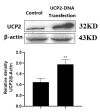
UCP2 expression was shown in both transfected and non-transfected cells. Each bar represents the mean ± SD of three independent experiments (** p < 0.01 vs. Control group).
Figure 6
Figure 6
Increasing UCP2 expression alleviated the cytotoxicity of AA I to HK-2 cells. (A) Inverted phase contrast microscope was used to observed the morphology of transfected or non-transfected cells after cells were treated with AA I and genipin (scale bar, 50 µM). (B) The nuclear morphology of transfected or non-transfected cells was evaluated using Hoechst 33,258 staining by fluorescence microscope (scale bar, 50 µM). The yellow arrows indicate the chromatin condensation, which is brightly stained and the red arrows show DNA fragmentation. (C) The intracellular ROS was measured using fluorescence microscopy (scale bar, 50 µM) with probe DCFH-DA.
Figure 7
Figure 7
MTT was used for cell viability and cytotoxicity. (A) HK-2 cells with or without UCP 2-DNA transfection were treated with different concentration of AA I for 24 h. (B) HK-2 cells with or without UCP2-DNA transfection were treated with different concentration of AA I + genipin for 24 h. Each bar represents the mean ± SD of three independent experiments (* p < 0.05; ** p < 0.01; vs. UCP2-DNA Transfection (−) Control group; # p < 0.05 vs. UCP2-DNA Transfection (+) Control group; & p < 0.05).
Figure 8
Figure 8
Increasing UCP2 expression alleviated AAI-induced apoptosis and oxidative stress. HK-2 cells with or without UCP2-DNA transfection were treated with 40 μM AA I + 25 μM genipin for 24 h. Caspase-3 activity (A), LDH activity (B), MDA generation (C) and GSH-Px activity (D) were detected on transfected or non-transfected cells. Each bar represents the mean ± SD of three independent experiments (* p < 0.05; ** p < 0.01; *** p < 0.001 vs. UCP2-DNA Transfection (−) Control group; # p < 0.05; ## p < 0.01 vs. UCP2-DNA Transfection (+) Control group; & p < 0.05; && p < 0.01).

References

    1. Balachandran P., Wei F., Lin R.C., Khan I.A., Pasco D.S. Structure activity relationships of aristolochic acid analogues: Toxicity in cultured renal epithelial cells. Kidney Int. 2005;67:1797–1805. doi: 10.1111/j.1523-1755.2005.00277.x. - DOI - PubMed
    1. Yang H.-Y., Chen P.-C., Wang J.-D. Chinese Herbs Containing Aristolochic Acid Associated with Renal Failure and Urothelial Carcinoma: A Review from Epidemiologic Observations to Causal Inference. BioMed Res. Int. 2014;2014:569325. doi: 10.1155/2014/569325. - DOI - PMC - PubMed
    1. Bastek H., Zubel T., Stemmer K., Mangerich A., Beneke S., Dietrich D.R. Comparison of Aristolochic acid I derived DNA adduct levels in human renal toxicity models. Toxicology. 2019;420:29–38. doi: 10.1016/j.tox.2019.03.013. - DOI - PubMed
    1. Rebhan K., Ertl I.E., Shariat S.F., Grollman A.P., Rosenquist T. Aristolochic acid and its effect on different cancers in uro-oncology. Curr. Opin. Urol. 2020;30:689–695. doi: 10.1097/MOU.0000000000000806. - DOI - PubMed
    1. Yeh Y.H., Lee Y.T., Hsieh H.S., Hwang D.F. Short-term toxicity of aristolochic acid, aristolochic acid-I and aristolochic acid-II in rats. Food Chem. Toxicol. 2008;46:1157–1163. doi: 10.1016/j.fct.2007.11.015. - DOI - PubMed

LinkOut - more resources