. 2022 Mar 28;23(7):3697.
doi: 10.3390/ijms23073697.

7-Ketocholesterol-Induced Micro-RNA-107-5p Increases Number and Activity of Osteoclasts by Targeting MKP1

Affiliations

7-Ketocholesterol-Induced Micro-RNA-107-5p Increases Number and Activity of Osteoclasts by Targeting MKP1

Guoen Li et al. Int J Mol Sci. .

Abstract

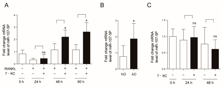
Osteoclasts (OCs), which are responsible for bone resorption, play a critical role in cholesterol-induced bone loss and recent studies have suggested that various micro-RNAs (miRs) contribute to modulating OCs. We hypothesized that 7-ketocholesterol (7-KC), a metabolite responsible for cholesterol-induced bone loss, induces miR-107-5p, which affects OCs. Overexpression and knock-down of miR-107-5p were performed using miR-107-5p mimic and anti-miR-107-5p, respectively. The effects of miR-107-5p on OCs were analyzed by tartrate-resistant alkaline phosphatase staining, qPCR, and Western blot. MiR-107-5p was upregulated after 7-KC exposure in receptor activator of nuclear factor kappa-Β ligand-stimulated OCs. Furthermore, miR-107-5p upregulation was also observed in tibiae from an atherogenic diet-fed mice compared with mice fed with a normal diet. MiR-107-5p overexpression enhanced the area and number of OCs, whereas inhibiting the endogenous expression of miR-107-5p generated by 7-KC had the opposite effect. Among the possible candidates, mitogen-activated protein kinase phosphatase-1, a stress-responsive dual-specificity phosphatase that inactivates mitogen-activated protein kinase (MKP1), has been proven to be a target gene of miR-107-5p, as demonstrated by the direct interaction between miR-107-5p and the 3'-untranslated region of MKP1. Collectively, our findings demonstrate that 7-KC-induced miR-107-5p promotes differentiation and function of OCs by downregulating MKP1.

Keywords: 7-ketocholesterol; micro-RNA-107-5p; mitogen-activated protein kinase phosphatase 1; osteoclast.


Conflict of interest statement

The authors state that they have no conflict of interest. The funders had no role in the design of the study, analysis or interpretation of data, writing of the manuscript, or decision to publish the results.

Figures

Figure 1
Figure 1
Atherogenic diet (AD) upregulates micro-RNA-107-5p (miR-107-5p). (A) Bone marrow-derived macrophages (BMMs) were prepared and incubated with or without 7-ketocholesterol (7-KC, 7 μM) in the presence of macrophage colony stimulating factor (M-CSF, 30 ng/mL) and receptor activator of nuclear factor kappa-Β ligand (RANKL, 40 ng/mL) for the indicated times, and then analyzed by qPCR to quantify the expression of miR-107-5p (n = 3–5). (B) Tibiae from mice fed with an AD or normal diet (ND) for 12 weeks were analyzed by qPCR to quantify the expression of miR-107-5p (AD, n = 8; ND, n = 8). (C) Primary osteoblasts were treated with 7-KC (7 μM) for the indicated time points and analyzed by qPCR to quantify miR-107-5p expression (n = 4). * p < 0.05 compared with each corresponding group; ns, not significant. Similar results were obtained in three independent experiments.
Figure 2
Figure 2
The expression level of miR-107-5p regulates osteoclast (OC) differentiation. BMMs were prepared, incubated with M-CSF (30 ng/mL) and RANKL (40 ng/mL) for 48 h, and transfected with 30 nM of miR-107-5p mimic or con mimic (AD). BMMs were prepared, incubated with M-CSF (30 ng/mL), RANKL (40 ng/mL), and 7-KC (7 μM) for 48 h, and transfected with 30 nM of anti-miR-107-5p or con inh (EG). After the indicated time points, total RNA was analyzed by qPCR to quantify the expression of miR-107-5p. The expression levels of BMM (A,E) were set to 1. Cells were incubated for 17 h and fixed to analyze tartrate-resistant acid phosphatase (TRAP)-positive OCs (B,F) and the expression of TRAP, nuclear factor of activated T cells 1 (NFAT2), calcitonin receptor (CTR), ATP6v0d2, and dendrocyte expressed seven transmembrane protein (DC-STAMP) were quantified by qPCR (C,G). More than 100 TRAP-positive multinucleated cells (MNCs) in each culture were randomly selected. The area (bold line) of the formed OCs was then measured. Representative photos of OCs. Scale bar; 200 μm (B,F). Mature OCs were loaded on whole dentine and transfected with miR-107-5p mimic or con mimic for another 5 d in the presence of M-CSF and RANKL. After TRAP staining, the cells were removed, and the slices were stained with toluidine blue. Representative photos of TRAP-positive OCs and resorption pits are shown (scale bar: 50 μm). The total pit area/number of TRAP-positive OCs was calculated (D). (A-G; n = 3 each group). * p < 0.05; ** p < 0.01; *** p < 0.001 compared with each corresponding control. Similar results were obtained in three independent experiments.
Figure 3
Figure 3
Identification of target for 7-KC-induced miR-107-5p in OCs. (A,D) BMMs were prepared, incubated with M-CSF (30 ng/mL) and RANKL (40 ng/mL) for 48 h, and transfected with 30 nM of miR-107-5p mimic or con mimic (A). BMMs were incubated with M-CSF, RANKL, and 7-KC (7 μM) for 48 h and transfected with 30 nM of anti-miR-107-5p or con inh. (D). Cells were incubated for 17 h and total RNA was analyzed by qPCR to quantify the expression of MAP kinase phosphatase 1 (MKP1), Src homology region 2 domain containing phosphatase-1 (SHP1), and phosphatase and tensin homolog deleted on chromosome 10 (PTEN). The expression levels with con mimic treatment (A) or con inh treatment (D) were set to 1. Cell lysates were subjected to Western blot analysis with antibodies against MKP1, SHP1, and PTEN. Antibodies against β-actin were used for normalization (n = 4). (B) BMMs were incubated with M-CSF, RANKL, and 7-KC. After the indicated time points, total RNA was analyzed by qPCR to quantify the expression of MKP1. After 65 h, cell lysates were subjected to Western blot analysis with anti-MKP1 Ab (n = 3). (C) The total RNA and tissue lysates of tibiae from AD- or ND-fed mice were analyzed by qPCR to quantify the expression of MKP1 and were subjected to Western blot analysis with antibodies against MKP1 (AD, n = 8; ND, n = 8). (E) BMMs were incubated with M-CSF, RANKL, and 7-KC for 48 h, and transfected with 30 nM of scRNA or si-MKP1 with 7-KC in the presence of M-CSF and RANKL. The cells were incubated for 17 h and analyzed to measure TRAP-positive MNCs. Representative photos of TRAP-positive OCs are shown (scale bar: 200 μm). The silencing of MKP1 by siRNA was confirmed by RT-PCR and qPCR (n = 3). * p < 0.05; ** p < 0.01; *** p < 0.001 compared with each corresponding control; ns, not significant. Similar results were obtained from three independent experiments.
Figure 4
Figure 4
MiR-107-5p specifically targets MKP1 by binding to the 3′-untranslated region (UTR) of MKP1. (A) The target site of miR-107-5p in 3′-UTR of MKP1 is conserved in humans and rodents (indicated with asterisks). (B) The 3′-UTR mutants of MKP1 are identical to the mouse 3′-UTR wild type (WT) of MKP1 except that it contains five single base substitutions (underlined in the figure) to disrupt pairing with mature miR-miR-107-5p (mmu-miR-107-5p). (C) The mouse 3′-UTR WT of MKP1 containing miR-107-5p binding sites or its mutated counterpart were cloned into a luciferase reporter vector and transfected into RAW264.7 cells with miR-107-5p mimic or anti-miR-107-5p with each corresponding control (n = 6). Luciferase assays were performed using a dual-luciferase reporter assay system. ** p < 0.01; *** p < 0.001 compared to its corresponding control; ns, not significant. Similar results were obtained in three independent experiments.

Similar articles

Cited by

References

    1. Orozco P. Atherogenic lipid profile and elevated lipoprotein (a) are associated with lower bone mineral density in early postmenopausal overweight women. Eur. J. Epidemiol. 2004;19:1105–1112. doi: 10.1007/s10654-004-1706-8. - DOI - PubMed
    1. Yamaguchi T., Sugimoto T., Yano S., Yamauchi M., Sowa H., Chen Q., Chihara K. Plasma Lipids and Osteoporosis in Postmenopausal Women. Endocr. J. 2002;49:211–217. doi: 10.1507/endocrj.49.211. - DOI - PubMed
    1. Pelton K., Krieder J., Joiner D., Freeman M.R., Goldstein S.A., Solomon K.R. Hypercholesterolemia promotes an oste-oporotic phenotype. Am. J. Pathol. 2012;181:928–936. doi: 10.1016/j.ajpath.2012.05.034. - DOI - PMC - PubMed
    1. Larsson B., Sundh D., Mellström D., Axelsson K., Nilsson A., Lorentzon M. Association between cortical bone micro-structure and statin use in older women. J. Clin. Endocrinol. Metab. 2019;104:250–257. doi: 10.1210/jc.2018-02054. - DOI - PubMed
    1. Oxlund H., Andreassen T.T. Simvastatin treatment partially prevents ovariectomy-induced bone loss while increasing cortical bone formation. Bone. 2004;34:609–618. doi: 10.1016/j.bone.2003.12.014. - DOI - PubMed

LinkOut - more resources